Chapter 2: Effective Use of Your Dive Computer

“Divers are surprised when symptoms of DCS develop after dives that appeared safe according to their dive computers. Remember, models reflect an average diver, not you.”

In recent years, dive computers have supplanted dive tables as the primary means of regulating dive profiles. Dive computers offer an advantage in that they enable the diver to dynamically establish different compartments as the controlling compartment, as conditions change during a dive. In reality, the compartments in a dive computer’s modeling software do not have to represent any particular tissue, as long as the guidance provided by the model results in an acceptable outcome — specifically, very little DCS.

In this chapter, you’ll learn:


Important Cautions

While the guidance provided by decompression models can be very useful, it is important for divers to keep in mind that dive schedules — whether they are presented in printed tables or on the screen of a dive computer — are limited in what they measure and in the assumptions upon which the model was constructed. Tissue compartment parameters can be adjusted, or new compartments can be added to an algorithm, if experience shows deficiencies in a given model — but in real time, the calculations are limited by the variables that are being processed. Algorithms can estimate limits based on time and pressure (depth) profiles for a given breathing gas, but they are not able to compute the impact of myriad real-time factors, including thermal status, exercise intensity, joint forces and a host of individual predispositions that are currently not well understood, let alone quantifiable in their impact on decompression stress.

Divers are often surprised when symptoms of DCS develop after dives that were conducted within the limits of their dive computers. It is important to remember, though, that while mathematical models predict outcomes, they do not guarantee them. The fact that a dive was conducted within the limits suggested by a dive computer (or a dive table) does not make a DCS hit “undeserved.” The mathematical algorithms provide guidance that must be evaluated and tempered by a thoughtful diver.

Many divers are also unaware of the fact that dive computers make use of many different mathematical models, or versions of different models; there is no universal standard. A single manufacturer may even use more than one model, possibly in a single type of computer. This makes it extremely difficult to assess the nuances of every system.


Basic Guidelines

There are some basic guidelines that can help to ensure the safe and effective use of a dive computer. The following considerations are intended to offer a somewhat light-hearted insight into what your dive computer can — and cannot — do.

It is helpful to think of your dive computer in these ways:

  • As a business competitor: Master it by learning its strengths and weaknesses.
  • As a date: It must be turned on for the relationship to work.
  • As a buddy: It should descend and ascend whenever, but only when, you do.
  • As a personal assistant: It reminds you of rules and schedules you might otherwise forget.
  • As an actor: It recites the lines without having to understand their implications.
  • As a politician: Do not believe everything it tells you.
  • As a hotel concierge: It will help you do what you want — but at a price.
  • As a stranger: It knows virtually nothing about your personal reality.
  • As a mate: Is it compatible with your friends?
  • As a news reporter: It will air your dirty laundry.
  • As a tool: Use it appropriately.

Specific Tips and Tricks

Push the Right Buttons

blank

You should know not only which buttons to push to make your computer work, but also which mathematical model or model derivation it employs in making its decompression computations. There is a surprising range in models, from conservative to liberal, and these differences may not be evident at first glance. For example, a computer may establish conservative limits for an initial dive but liberal limits for repetitive diving. It is best to learn enough about the various available models and derivations before you select a dive computer, so you are sure to choose one that is compatible with your own level of risk tolerance. Choosing one purely based on familiarity may not be the best strategy. Even if you have had good outcomes on previous dives with a computer, it does not guarantee that it will be the best one for your future diving. Accumulating knowledge takes commitment, but informed planning for decompression safety should be a top concern.

Tune In and Turn On

Failing to turn on your dive computer (or to take it with you on a dive) may sound like a joke, but it does happen and can create real problems. No computer can factor in the exposure profile of a previous dive if it was not there. And any decompression model is invalid unless you start using it when you are “clean” — fully off-gassed from any previous dives. If you forget to take your computer with you on a dive early in a repetitive series, you are then restricted to using tables for the duration of that series (assuming that you are able to manually compute the exposure of the unmonitored dive). And do not even think about hanging your computer on a downline during a surface interval in an effort to compensate for having forgotten it on an earlier dive; there may be stories about that happening, but it is not a responsible practice.

Use It Appropriately

The only person who does not have to worry about taking a dive computer on every dive is the one who uses it solely as a datalogger — that is, only to record time and depth information instead of to calculate decompression profiles. Remember, however, that using your computer simply to log your time and depth data means that you must still plan all your dives using dive tables and must recompute your repetitive group status afterward, as appropriate. You cannot move in and out of relying on your computer’s decompression computations unless it has recorded all of your exposure profiles.

Remember Its Limitations

Dive computers are wonderful at carrying out programmed mathematical computations, but they are blind to the many insights you may have before, during and between your dives. For example, your dive computer knows nothing about your personal health status, your level of physical fitness or your individual susceptibility to decompression stress. It also knows nothing about your thermal stress or physical efforts during or between dives. The fact that many dive computers display water temperature might suggest that thermal stress is factored into the device’s algorithms. A water temperature reading, however, provides no useful information regarding thermal stress, since the diver carrying the device could be wearing anything from a bathing suit to a wetsuit without a hood to a cold-water drysuit with a hood, gloves and cold-water undergarments. More important, it is not yet possible to directly compute the impact of differences in thermal status during different parts of a dive, even if the computer was able to measure the diver’s core temperature and skin temperature in key spots.

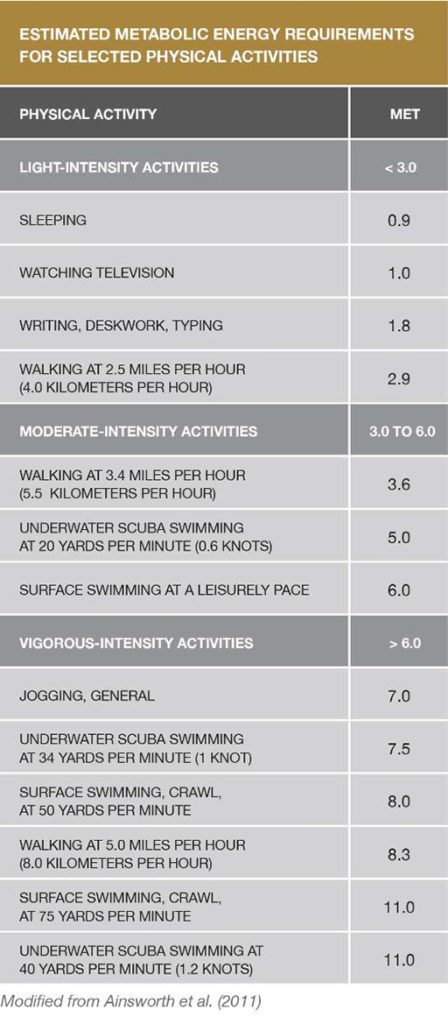
We do know that being warm (rather than cool or cold) during the compression and bottom phase of a dive promotes inert gas uptake (not optimal), and that being warm during the decompression phase promotes elimination (optimal). While impractical for the comfort-loving diver, decompression safety is optimized by being neutral or cool during the inert gas uptake phase of descent and bottom time and warm during the inert gas elimination phase of ascent. While the concept of thermal changes on decompression stress is clear, we are still years away from being able to quantify the real-world effects of these factors for dive-planning purposes. Similarly, while some computers are able to track gas consumption, we have much to learn before this information can be meaningfully incorporated into decompression models. Variations in air consumption can reflect differences in the depth of a dive or in the diver’s experience, level of anxiety or degree of physical exertion. The bottom line is that interpreting the precise physiological impact of the interactions among these diverse factors is exceedingly difficult, requiring thoughtful practice by divers.

Heed Your Computer’s Readings

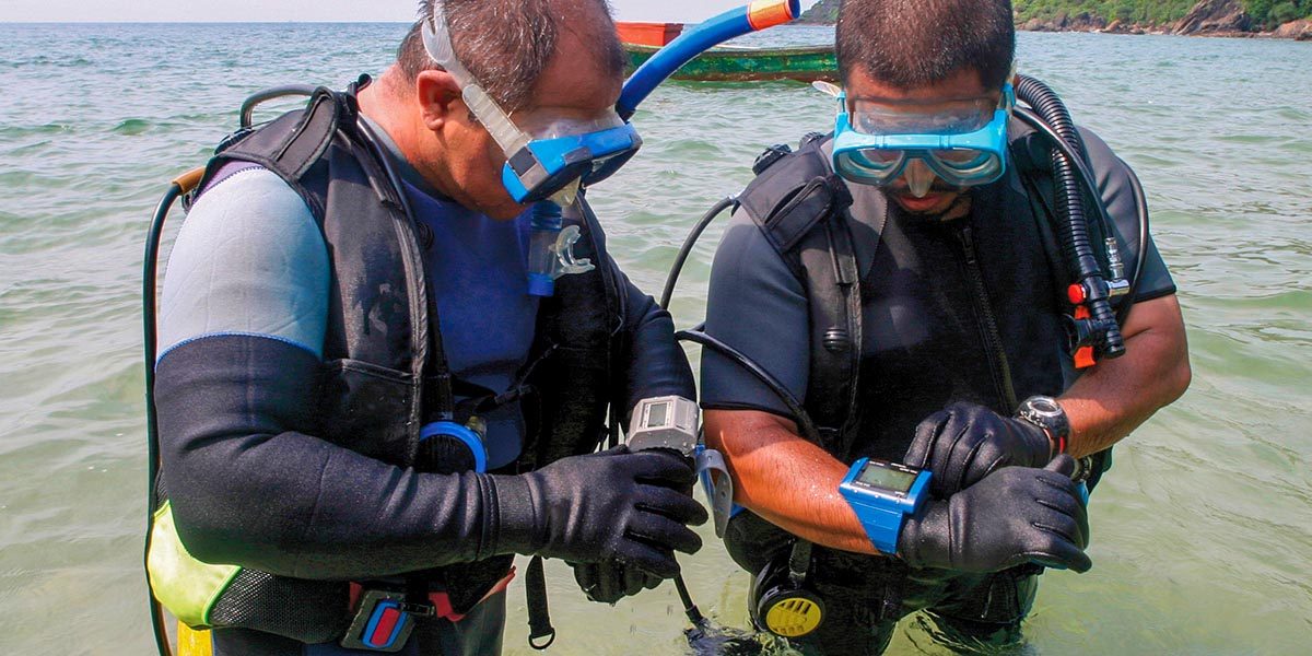
Divers need to pay attention to their dive computers if the information provided is to be of any use. Be aware that confirmation bias can promote risky behavior. “Getting away with” a risky exposure once, twice or even many times may eventually catch up with you. It may not truly be safe for you or for a partner who might have a higher degree of susceptibility to decompression stress. Those who wish to worry less about their exposure will have greater peace of mind if they choose a computer that employs an extremely conservative decompression model. It is also important to pay attention to your dive computer. If you are diving with a group, do not forget that there can be considerable variability in the guidance provided by different computers or computers with different user-selected settings. That means there is considerable benefit in diving with others who use a computer with a similar decompression model and settings, because if modest discrepancies arise, following the most conservative directive will likely not be terribly burdensome for the group. But if members of a group are using dive computers with substantially different models, and each diver wishes to follow his or her own device, it can lead to a breakdown in the buddy system.

Do Not Rely Blindly On Your Computer

Although heeding your computer is important, do not take its advice unthinkingly. The same profile can sometimes be conducted without problem again and again, right up to the dive where it does not prove safe. Divers often try to blame a specific factor, such as dehydration, for the development of symptoms following one dive but not another. This approach is not productive. The range of variables in play during a dive are rarely identical, and there is a probabilistic element to decompression risk — that is, chance can play a role in the manifestation of DCS.

The best approach is to avoid the extremes of either fatalistic resignation or smug focus on a single supposed magic bullet. There are many, many small steps you can take to make any dive safer. The most important one is to stay within a reasonably conservative time-depth profile and to add safety stops to every dive. Other important steps are to minimize your exercise intensity and avoid overheating during the gas-uptake phase of your dive, to choose the right breathing gas, to practice enough that you are able to perfectly control your buoyancy, to remain well-rested and well-hydrated, choose more conservative user-adjustable settings on the computer, and to dive with a partner who has similar goals and follows similar practices. Adding small safety margins to each step can help to provide a comfortable security cushion. Dive computers are powerful tools, but sound knowledge of diving physiology, good physical conditioning and adherence to thoughtful practices offer the best protection for divers.

Keep It With You

If you do develop DCS symptoms, you should keep your computer with you when you go for medical evaluation. Some facilities may have the ability to download or review your profile to aid in the evaluation of your case. The medical staff will surely appreciate seeing confirmation of your description of the events that precipitated your symptoms.

Next: Chapter 3 – Diagnosing Decompression Sickness >

Chapter 3: Diagnosing Decompression Sickness

blank

“While DCS is commonly thought of as a bubble disease, bubbles are probably only the gateway to a complex array of consequences and effects.”

DCS may develop when a diver’s degree of supersaturation is so high (or, stated another way, if the elimination gradient is so steep) that a controlled transfer of inert gases from the body’s tissues to the bloodstream — and then from the bloodstream to the lungs and the lungs to the environment — is not possible. If that removal process is inadequate, inert gases will come out of solution and form bubbles that can distort tissues, obstruct blood flow, cause mechanical damage (to the joints, for example) and/or trigger a cascade of biochemical responses.

Although much is known about DCS, its mechanisms of insult are still being investigated. And while DCS is commonly thought of as a bubble disease, bubbles are probably only the gateway to a complex array of consequences and effects.

In this chapter, you’ll learn about:


blank
Skin mottling like this is characteristic of cutis marmorata, a condition that can warn of likely development of more serious Type 2 symptoms.

Signs and Symptoms of DCS

The collective insult to the body’s systems can produce symptomatic DCS. The condition’s primary effects may be evident in the tissues that are directly insulted. Its secondary effects can compromise the function of a broad range of tissues, further jeopardizing the diver’s health.

The ability to recognize the signs, or objective evidence, and the symptoms, or subjective perceptions, of DCS — and to differentiate them from signs and symptoms less likely to be associated with DCS — is important. A variety of classification systems have been established for DCS. One common approach is to describe cases as Type 1 or Type 2.

Type 1 DCS

Type 1 DCS is usually characterized by musculoskeletal pain and mild cutaneous, or skin, symptoms. Common Type 1 skin manifestations include itching and mild rashes (as distinct from a clear mottled or marbled and sometimes raised discoloration of the skin — a condition that is known as cutis marmorata that may presage the development of the more serious symptoms of Type 2 DCS). Less common but still associated with Type 1 DCS is obstruction of the lymphatic system, which can result in swelling and localized pain in the tissues surrounding the lymph nodes — such as in the armpits, groin or behind the ears.

blank
Collage showing pain at several part of body

The symptoms of Type 1 DCS can build in intensity. For example, pain may originate as a mild ache in the vicinity of a joint or muscle and then increase in magnitude. However, the pain associated with DCS does not typically increase upon movement of the affected joint, although holding the limb in one position rather than another may reduce discomfort. Such pain can ultimately be quite severe.

Type 2 DCS

blank
The Romberg test evaluates postural control. The sharpened Romberg, which includes crossing the arms and putting one foot in front of the other, is more sensitive to changes in static balance.

Type 2 symptoms are considered more serious. They typically fall into three categories: neurological, inner ear and cardiopulmonary. Neurological symptoms may include numbness; paresthesia, or an altered sensation, such as tingling; muscle weakness; an impaired gait, or difficulty walking; problems with physical coordination or bladder control; paralysis; or a change in mental status, such as confusion or lack of alertness. Inner-ear symptoms may include ringing in the ears, known as “tinnitus”; hearing loss; vertigo or dizziness; nausea; vomiting; and impaired balance. Cardiopulmonary symptoms, known commonly as “the chokes,” include a dry cough; chest pain behind the sternum, or breastbone; and breathing difficulty, also known as “dyspnea.” The respiratory complaints, which are typically due to high bubble loads in the lungs, can compromise the lungs’ ability to function — threatening the affected diver’s health, and even life, if treatment is not sought promptly.

Type 2 symptoms can develop either quickly or slowly. A slow build can actually obscure the seriousness of the situation, by allowing denial to persist. For example, fatigue and weakness are common enough concerns, especially if their onset is protracted, that they can be very easy to ignore. Less common symptoms, such as difficulty walking, urinating, hearing or seeing — especially if their onset is quick — can sometimes prompt faster recognition of the existence of a problem. It is fair to say that divers can initially be reluctant to report symptoms, though they usually will do so if their symptoms do not go away. This is a shortcoming divers should be aware of, lest they fall prey to it.

Presentation of DCS

The presentation of DCS is frequently idiosyncratic — that is, its “typical” pattern can be atypicality. In some cases, an affected diver’s chief complaint may draw attention away from more subtle but potentially more important symptoms. The following list ranks the initial manifestations of DCS, from those most commonly to least commonly reported (Vann et al. 2011):

  • Pain, particularly near the joints
  • Numbness or paresthesia
  • Constitutional concerns — such as headache, lightheadedness, unexplained fatigue, malaise, nausea and/or vomiting, or anorexia
  • Dizziness or vertigo
  • Motor weakness
  • Cutaneous, or skin, problems — such as an itch, rash, or mottling (“cutis marmorata”)
  • Muscle discomfort
  • Impaired mental status
  • Pulmonary problems — such as breathing difficulties (“the chokes”)
  • Impaired coordination
  • Reduced level of consciousness
  • Auditory symptoms — such as hearing sounds that are not there or having a hard time hearing
  • Lymphatic concerns — such as regional swelling
  • Bladder or bowel dysfunction — such as retention of urine
  • Compromised cardiovascular function

According to this recent review, pain and numbness, also known as paresthesia, were reported initially in nearly two-thirds of cases of DCS, constitutional symptoms in approximately 40 percent of cases, dizziness/vertigo and motor weakness in approximately 20 percent, and cutaneous symptoms in approximately 10 percent (Vann et al. 2011).


Differential Diagnosis of DCS

blank

DCS is a high-profile diving injury because of its potential severity. But divers need to remember that not all diving-related problems turn out to be DCS. When two or more conditions have overlapping symptoms, as is the case with many diving-related injuries, differential diagnosis is the process by which medical personnel figure out which of the potential conditions is most likely responsible for the symptoms.

The term decompression illness (DCI) was coined to encompass both DCS and the related condition known as arterial gas embolism (AGE), the latter arising from barotrauma of the lungs that introduces gas into the systemic bloodstream. Some of the other conditions and circumstances that involve similar symptoms include inner-ear barotrauma; middle-ear or maxillary sinus overinflation; contaminated breathing gas; oxygen toxicity; musculoskeletal strains or trauma sustained before, during or after a dive; marine life envenomation; immersion pulmonary edema; water aspiration; and coincidental neurological disorders, such as stroke (Vann et al. 2011). Thermal stress — sometimes due to excessive heat, but usually due to cold exposure — can also be responsible for similar symptoms. In some cases, a careful medical history can easily rule out one diagnosis or another. For example, symptoms of immersion pulmonary edema often develop at depth. In such a case, a good history would rule out DCS, which only develops after significant decompression stress during ascent.

It is essential for divers with any of these symptoms to seek medical evaluation and support. While first responders are able to perform initial analysis of an injured individual, such as administering a field neurological assessment, the capabilities of nonphysicians do not come close to the clinical skills and insights held by experienced clinical specialists.

Next: Chapter 4 – Treating Decompression Sickness >

Chapter 4: Treating Decompression Sickness

blank

“If signs or symptoms consistent with DCS develop, initiate appropriate first aid and contact the nearest emergency medical services. For additional emergency assistance contact DAN +1-919-684-9111.”

There are several elements to the effective management of DCS, specifically on-the-scene evaluation and first aid, transport and definitive medical evaluation and treatment. Anyone who has suffered DCS should seek appropriate evaluation, and possibly ongoing care, from a physician well informed about diving-related medical issues.

In this chapter, you’ll learn about:


On-the-Scene First Aid

The foundation of first aid is basic life support. The primary first aid measure for DCS is delivery of supplemental oxygen in the highest concentration, or fraction, that is practical (Longphre et al. 2007). High oxygen fractions, if provided rapidly and over a sustained period, can reduce or even eliminate symptoms of DCS, albeit often only temporarily if definitive treatment is not secured. Continuous-flow oxygen systems, using non-rebreather or pocket masks, are frequently available in diving environs; however, such equipment delivers modest oxygen fractions. Much higher fractions can be achieved with demand masks, though they are appropriate only for conscious individuals able to breathe on their own.

blank
A variety of different masks can be used with oxygen systems.
blank
DAN experts developed this remote emergency oxygen (REMO2) surface oxygen rebreather for first-aid use (Pollock and Natoli 2007).

Rebreather systems are another on-the-scene option; such systems permit the unused oxygen in the diver’s exhalations to be recycled, or rebreathed. A rebreather apparatus can thus provide high fractions with minimal gas use and may prove especially helpful in settings where the supply of oxygen is limited (Pollock 2004; Pollock and Natoli 2007).

Chemical oxygen generating systems — devices with a long shelf life that deliver oxygen via a chemical reaction — may in some situations be the only option available. However, if emergency medical services are not readily accessible, such devices are unlikely to provide a sufficient oxygen supply (Pollock and Natoli 2010).


Subsequent Evaluation

First aid is just the first step in treating an affected diver. Anyone who has experienced symptoms associated with DCS is advised to seek subsequent medical evaluation. This should occur even if the diver’s symptoms improved or disappeared upon the administration of oxygen, since subtle issues can be missed or symptoms can return once oxygen delivery is stopped. For the same reason, it is advisable to seek input from an experienced dive-medicine specialist — someone aware of all the nuances in the presentation, course and treatment of DCS.


Hyperbaric Oxygen Therapy

The definitive treatment for DCS is hyperbaric oxygen (HBO) therapy, or the delivery of pure oxygen at a pressure substantially higher than that of atmospheric pressure. HBO therapy reduces the size of any bubbles and improves gradients which promote oxygen delivery and inert gas elimination. HBO therapy is typically delivered in recompression chambers.

blank
This is a monoplace hyperbaric chamber — able to hold a single patient, without any inside support personnel, or “tenders.”

A common HBO regimen is the U.S. Navy Treatment Table 6 (USN 2008). According to this regimen, the hyperbaric chamber is initially pressurized to 2.8 atmospheres absolute (ATA), equivalent to the pressure found at 60 feet (18 meters) of seawater. The patient breathes pure oxygen, interspersed with scheduled periods of breathing regular air to reduce the risk of oxygen toxicity. The usual duration of the USN TT6 treatment is just under five hours, but extensions can be added as required, based on the patient’s response.

blank
This is a small multiplace and multilock hyperbaric chamber; it can hold multiple patients plus inside tenders. Personnel or equipment can be transferred into or out of the chamber while treatment is ongoing.

HBO treatment can be conducted in a monoplace chamber, often an acrylic tube sized to hold just one patient, or in a multiplace chamber, sized to accommodate one or more patients plus one or more “tenders” — that is, technicians or other medical personnel. Multilock chambers are designed to allow patients, tenders or equipment to be transferred into and out of the chamber while treatment is ongoing.

The course of HBO therapy will vary according to the particulars of each case; both the presentation of DCS and its response to treatment can be idiosyncratic. A full resolution of DCS symptoms can often be achieved with one or sometimes multiple HBO treatments. In some cases, however, resolution will be incomplete, even after many treatments. The normal clinical approach is to continue the treatments until no further improvement is seen in the patient’s symptoms. Modest residual symptoms will then often resolve slowly, after the treatment series is ended. Full resolution of symptoms can sometimes take months to achieve and in some instances may never be realized.

blank
This is a large multiplace and multilock hyperbaric chamber, in which multiple pressure exposures can be conducted simultaneously.

In-Water Recompression

blank

In-water recompression may be an alternative to chamber recompression in remote locations, if there is neither a nearby chamber nor the means to quickly transport the patient to a chamber elsewhere. The technique involves bringing the diver underwater again, to drive gas bubbles back into solution to reduce symptoms and then slowly decompress in a way that maintains an orderly elimination of the excess gas.

While in-water recompression is simple in concept, it is practical only with a substantial amount of planning, support, equipment and personnel; appropriate water conditions; and suitable patient status. Critical challenges can arise due to changes in the patient’s consciousness, oxygen toxicity, gas supply, and even thermal stress. An unsuccessful in-water recompression may leave the patient in worse shape than had the attempt not been made. The medical and research communities are divided on the utility of in-water recompression. It is beyond the scope of this publication to consider all of the relevant factors, but it is fair to say that there are probably more situations when in-water recompression should not be undertaken than situations when it would be a reasonable choice.

As a general rule, a diver who develops symptoms consistent with DCS should be removed from the water, and first aid should be delivered on the surface, even if there is likely to be a delay before definitive medical care can be sought.


Emergency Resources

The best course of action, if signs or symptoms consistent with DCS (or any other serious injury) develop, is to initiate appropriate first aid and then immediately contact the nearest emergency medical services (EMS). The next step should be to contact DAN to seek advice regarding the proper progression of care. The organization’s emergency hotline number is +1-919-684-9111.

It is generally not appropriate to show up unannounced at the nearest hyperbaric chamber. This could mean bypassing a facility where the victim might be able to receive a more thorough and appropriate evaluation. Remember that not all injuries associated with diving are DCS, even if it seems so in the heat of the moment. In addition, the chambers at some facilities are not available to treat divers at all times or ever. One of the challenges within North America is the shrinking number of hyperbaric chambers that accept diving casualties, particularly outside normal business hours.

The key point to remember is that establishing contact with emergency medical services and DAN can ensure timely and appropriate case management. When in doubt, call.

WHEN YOU CALL THE
DAN EMERGENCY HOTLINE

  1. Tell the operator you have a dive emergency. The operator will confirm your name, location and phone number, and ether connect you directly with DAN medical staff or have someone call you back at the earliest possible moment.
  2. The medical staff member may make an immediate recommendation or call you back after making arrangements with a local physician
  3. The medical staff member may ask you to wait by the phone while arrangements are being made. These plans may take 30 minutes or longer, as complex coordination is often required. If the situation is life-threatening, arrange safe transport for the diver to the nearest medical facility for immediate stabilization and assessment first. Then call the DAN Emergency Hotline for consultation with the local medical provider.

Even if symptoms were not severe and they resolved completely, a diver who has had multiple bouts of DCS must take special considerations. Especially if DCS is recurring following otherwise safe dive profiles, a dive medical specialist must be consulted to determine if diving can be resumed safely.

DAN EMERGENCY HOTLINE +1 919-684-9111

Next: Chapter 5 – Factors Contributing to Decompression Stress >

Chapter 5: Factors Contributing to Decompression Stress

blank

“A number of factors contribute to your individual susceptibility to DCS and can even alter your susceptibility from day to day.”

The most significant risk factor is your exposure profile — that is, the time, depth and ascent rate of your dives. Some degree of exposure intensity is required to initiate a decompression insult, regardless of the presence of other predisposing factors.

There are a host of factors, however, that can play a role in your outcome if you experience an exposure sufficient to make DCS a possibility. Several common risk factors are outlined in this chapter.

In this chapter, you’ll learn about:


Workload

During the Dive

blank

The timing and intensity of exercise during a dive can substantially affect your risk of DCS. A high workload during the descent and bottom phase of a dive will increase your inert gas uptake, effectively increasing the subsequent decompression stress. And exertion near the end of or immediately after a dive, particularly if it involves high joint forces, can stimulate bubble formation and increase the likelihood of bubbles passing through the lungs without being filtered out of the circulation.

You should keep your exercise intensity as low as possible during the bottom phase of a dive. Mild exercise — on the order of no more than two to three times resting effort, and with very low joint forces — is appropriate during the upper ascent and stop phases of a dive. However, any exercise, particularly exercise involving high joint forces, should be avoided as long as possible after a dive. If you are unable to avoid postdive exercise, you should keep your dive profiles very conservative to minimize your overall risk.


Thermal Stress

A diver’s thermal status has long been known to influence decompression risk. The impact is best appreciated by considering the two fundamental phases of every dive: the descent and bottom phase, when gas uptake occurs, and the ascent and stop phase, when gas elimination occurs.

Two Phases

During the descent and bottom phase of a dive, a relatively warm state results in increased inert gas uptake; this is equivalent to conducting a deeper and/or longer dive. On the other hand, if you can maintain a cool or thermoneutral state during your descent and bottom phase, you will effectively reduce your inert gas uptake. This beneficial effect will be further magnified if you exert yourself as little as possible during this phase.

blank

During the ascent and stop phase of your dive, a relatively warm state will promote inert gas elimination, thus reducing overall decompression stress. On the other hand, a cool or cold state during this phase will reduce inert gas elimination, effectively prolonging and possibly increasing decompression stress.

The decompression hazard associated with hot water suits — which effectively establish a warm condition in both phases of a dive — was established in a study of North Sea divers conducted 30 years ago (Shields and Lee 1986). The impact of thermal status on decompression stress was even more elegantly demonstrated in a recent study conducted by the U.S. Navy (Gerth et al. 2007). The controlled conditions of a research study cannot be directly correlated with everyday diving practices, but the key message from these studies is the importance of thoughtful thermal status. Keeping neutral on your way down — certainly avoiding unnecessary overheating — and warm on your way up (approaching a cool-warm pattern) will reduce the risk of DCS in comparison to being warmer on your way down and cool on your way up (a warm-cool pattern).


Optimal Practices

The difficulty comes in reconciling optimal practices for decompression safety with divers’ desires and normal practices. It is understandable for divers to want to warm themselves before the start of a dive, in anticipation of getting colder as the dive proceeds. Historically, divers did this by pouring warm water into their wetsuits or gloves before a dive. Then some divers began to place chemical hot packs in their suits. Modern divers have even more choices available to them, due to today’s array of active heating garments suitable for use with either wetsuits or drysuits. The problem, though, remains the same: warming the body’s peripheral tissues enhances circulation and increases the delivery of inert gases, particularly if the heating is applied early in a dive, when inert gas uptake is typically at its highest level. Furthermore, both warm water and chemical hot packs lose their effectiveness over time, potentially creating the warm-cool pattern shown to generate the greatest risk of DCS. Even active heating garments — which are able to keep the diver warm throughout a dive — involve a somewhat elevated risk. As shown with hot water suits, a warm-warm pattern, while associated with less DCS than a warm-cool pattern, remains more hazardous than a cool-warm pattern. Practically, divers should maintain adequate thermal protection to ensure clear thinking and physical capability. Excessive warming during dives should be avoided.

Divers must also keep in mind that postdive warming can also influence decompression risk. Indulging in rapid postdive warming, such as by taking a hot shower or getting into a hot tub, decreases the solubility of inert gas in tissues. This will promote the formation of bubbles in local tissues, often before perfusion increases sufficiently to remove the gas. Skin symptoms, fortunately often mild and transient — not cutis marmorata — can develop with rapid warming of the skin postdive. The challenge is to get divers to prioritize safe decompression over pure comfort. If an active heating system is to be used, this means leaving it off or on its lowest setting during your descent and bottom phase, and then turning it up a modest amount during your ascent and stop phase. It also means delaying the postdive pleasure of jumping into a hot shower or hot tub. If delayed gratification is not your style, then you should use more conservative dive profiles to reduce your overall risk.


Postdive Air Travel

blank

Modern air travel has made distant dive locations easily accessible. Flying to a destination near sea level before diving engenders virtually no risk (outside the possibility of mild dehydration or impairment due to long periods of relative immobility). Since flights end with compression, the tissues of plane passengers will be undersaturated upon landing and subsequently accumulate inert gases to re-establish equilibrium with the ambient pressure.

Flying after diving, however, increases decompression stress, since the pressure in an aircraft cabin is lower than that of ground-level atmospheric pressure. Commercial aircraft must have the capability of maintaining cabin pressure at an equivalent of 8,000 feet (2,438 meters), approximately 0.76 ATA. This does not mean that cabin pressure is always maintained at higher pressures. A recent study found that 10 percent of the commercial flights tested had cabin pressures exceeding 8,000 ft (Hampson et al. 2013). Now imagine that you have just completed a dive to 66 feet (20 meters), where you experienced an underwater pressure of 3.0 ATA. Your return to the surface, and the 1.0 ATA pressure of sea level, has already subjected your body to a threefold reduction in pressure (3.0:1.0). If you then get on a plane that has a cabin altitude of 8,000 feet, you would be subjecting yourself to a fourfold reduction (3.0:0.76) and thus to even greater decompression stress. Furthermore, should your plane suffer an unlikely but not impossible cabin depressurization, you would be subjected to a much greater decompression stress.

DAN and the Undersea and Hyperbaric Medical Society (UHMS) held a workshop in 2002 to review the available data regarding the decompression stress of flying after diving and develop consensus guidelines (Sheffield and Vann 2004). There were two important stipulations regarding the guidelines: first, adhering to them will reduce your risk but offers no guarantee that you will avoid DCS, and second, observing even longer surface intervals than the recommended minimums will reduce your DCS risk further still. Keeping in mind these caveats, these are the guidelines:

  • After a single no-decompression dive, a minimum preflight surface interval of 12 hours is suggested.
  • After multiple dives per day or multiple days of diving, a minimum preflight surface interval of 18 hours is suggested.
  • After dives requiring decompression stops, there was little evidence on which to base a recommendation, but a preflight surface interval substantially longer than 18 hours is considered to be prudent.

There are two further factors of note regarding the DAN-UHMS flying after diving guidelines:

  • They apply to flights at altitudes of between 2,000 and 8,000 feet (610 and 2,438 meters). The effect of a flight at an altitude below 2,000 feet was considered mild enough not to warrant special consideration — giving divers the flexibility to engage in modest postdive air travel, such as a short, low-altitude, inter-island flight.
  • They apply only to divers who have experienced no DCS symptoms. It is essential that a diver who is experiencing any symptoms consistent with DCS seek treatment prior to flying.

It is important to remember that any postdive ascent to a higher altitude — even using ground transportation — increases your decompression stress. Taking a cautious approach in such a case, by keeping your final dive profiles more conservative and/or delaying your travel to the higher altitude, is always advisable. The U.S. Navy has generated detailed tables and procedures that allow computation of exposure limits to a greater range of altitudes and with more time flexibility than the DAN-UHMS guidelines (USN 2008). It is important to appreciate, though, that these are simply mathematical constructs based on the same data used in developing the DAN-UHMS guidelines. Furthermore, they require the computation of repetitive groups for planning, something that is done with dive tables but not dive computers. Despite these limitations, they can be useful, particularly for a regular pattern of altitude diving.


Medical and Physical Fitness

blank

Poor medical and physical fitness can compromise your safety in general and may increase your risk of DCS. Definitive data are limited, but there is no question that it is prudent to maintain a high level of physical fitness and to dive progressively more conservatively as your fitness level declines. Safe diving is possible throughout much of a normal life span, but it is important for all divers to seek regular, objective evaluation of their capabilities and to adapt their diving practices accordingly. But even for divers who have transitioned from independent to more dependent forms of diving, in which they increasingly rely on the support of others, there will ultimately be a point at which they should hang up their fins.

Physical Activity Recommendations

Adults need two types of regular activity to maintain or improve their health—aerobics and strength training. The Centers for Disease Control and Prevention’s 2008 Physical Activity Guidelines for Americans recommends at least two and a half hours a week of moderate-intensity aerobic exercise to achieve health benefits, and five hours a week to achieve additional fitness benefits. And just as important as engaging in aerobic exercise is doing muscle-strengthening activities at least two days a week.

While good health and physical fitness will not solve all problems, the foundation is an important one. An adequate physical reserve can allow a quick response to keep a small problem from becoming a serious one. Relevant scenarios can be easily imagined for almost any dive.

Regular aerobic exercise has many positive benefits. Cardiac reserve is the difference between the rate at which the heart pumps blood at rest and its maximum capacity. An increase in this reserve may make it easier to meet the physical demands of diving activity and stress. Blood values of cholesterol can improve, reducing susceptibility to heart disease. Insulin sensitivity can improve, reducing the risk of developing diabetes. While the data specific to diving are much more preliminary, there is also some evidence that higher levels of aerobic fitness may contribute to a reduced decompression stress.

Most individuals are aware that being fit can improve quality of life. A major problem, however, is that time takes a toll on us. The ease with which we maintain our fitness level in our 20s can be very different from the reality as decades pass. Aerobic fitness typically declines on an average of one percent per year after age 30. The important point is that while some decline may be unavoidable due to a gradual loss of muscle mass and a reduction in the metabolic activity of aging muscle, the rate can be slowed and the reserve range broadened by adopting healthy lifestyles as early as possible.

The physical fitness needed for diving will vary with the demands of the environment, the equipment, and the nature of the dive. The best strategy is to incorporate regular physical activity into your life to improve or preserve your capabilities, and to prolong your diving life. Do not count on diving to keep you physically fit. If done properly, it should be your relaxing time in the water. To maintain or build aerobic capacity and strength, swim, cycle, run, or do whatever other physical activities you can enjoy. The more fit you are, the longer you get to play.

Detailed physical activity recommendations can be found at cdc.gov/physicalactivity/everyone/guidelines.


State of Hydration

blank
water

Dehydration gets a substantial amount of attention in the lay diving community as a risk factor for DCS, but probably more than is warranted. Sound hydration is important for good health, both for general and for diving health, but for your dive profile, thermal stress and exertion level are far more important risk factors for DCS. The undue focus on dehydration is probably a result of two issues. The first is that substantial fluid shifts can result from DCS, often creating a need for substantial fluid therapy and creating an impression that this was a cause, rather than a consequence, of the disease. The second issue is human nature — the understandable desire to assign blame for a condition that is capricious. DCS is fickle. A diver may adhere to a similar dive profile many times without incident but then develop DCS while following the very same profile. It is comforting to try and identify a single causal agent, even if this is more wishful than factual. It is important for divers to realize that a multitude of factors can subtly affect the risk on any one dive and that there is a probabilistic nature to the disease. Focusing on a range of strategies to reduce risk is more effective than trying to put all the blame on one.


Breathing Gas Mixture

The particular breathing gas mixture you use, and how you use it, can play a role in the development of DCS. A mixture known as enriched air nitrox, or simply nitrox, is increasingly popular for recreational diving. The percentage of oxygen in the mix is increased, reducing the nitrogen fraction. This means that there is less nitrogen uptake at a given depth. The decompression effect of nitrox, compared to that of air, can be calculated by computing what is known as equivalent air depth (EAD). The risk of DCS when diving with nitrox to the EAD table limits is not appreciably different than diving with air to the air table limits. It is possible to achieve a decompression safety buffer by using nitrox with air table limits, since this will reduce your inert gas uptake compared to using air.

blank

The critical caveat with nitrox is that its higher oxygen content means that a diver breathing nitrox is at risk of developing oxygen toxicity at a shallower depth than a diver breathing air. The recommended maximum partial pressure of oxygen — partial pressure being the portion of the total gas pressure represented by a single gas — is 1.4 ATA for recreational diving. When diving with air (21 percent oxygen), this level is reached at a seawater depth of 187 feet (57 meters) — beyond the usual recreational diving limit (187 feet of seawater = 6.6 ATA * 0.21 ATA oxygen in air = 1.4 ATA). When diving with a 32 percent nitrox mixture, this level is reached at a seawater depth of 111 feet (34 meters), and with 36-percent nitrox at just 95 feet (29 meters) — depths commonly reached by recreational divers.


Carbon Dioxide Level

blank

Elevated levels of carbon dioxide can increase the risk of DCS and lower the threshold for oxygen toxicity. Carbon dioxide is a potent vasodilator, meaning it causes the blood vessels to widen, increasing blood flow and the delivery of gases to tissues. Factors that can raise divers’ carbon dioxide levels include the increased dead space of breathing equipment (gas volume that must be moved but does not take part in gas exchange), the additional work of breathing dense gas underwater, and exercise. Using a well-designed and well-maintained breathing system, minimizing physical effort and remaining relaxed while underwater can minimize carbon dioxide increase.


Patent Foramen Ovale

blank

Patent foramen ovale (PFO), literally, open ovale window, is a persistent opening between the left and right atria of the heart. In fetal circulation, a major opening between the atria allows blood to largely bypass the lungs that are not yet being used for gas exchange. A flap normally closes over the opening after birth and is sealed by tissue. In approximately 25 percent of the population, a partial opening remains, the PFO. The opening can range in size from functionally irrelevant to physiologically significant, the latter allowing a substantial portion of blood to be shunting from the right heart to the left heart, bypassing gas exchange and filtration in the lungs. PFOs typically produce no symptoms and individuals are unaware of their status unless they are incidentally discovered through medical tests. However, the presence of a large PFO may increase the risk of DCS in divers who develop significant bubble loads. The correlation between PFO and DCS risk is not a clear one, since the frequency of PFO in the population is fairly high while DCS is relatively rare. The safest strategy — even if you have not been diagnosed with a PFO, but most certainly if you have — is to dive in a manner calculated to keep your bubble load low; this effectively eliminates any concern that bubbles might pass through a PFO and bypass the lungs, where they would normally be filtered out.

The most commonly held consensus is that screening all divers for PFO is probably not warranted. And even in divers who have been diagnosed with a PFO, deciding whether it warrants surgical closure is a choice that each individual should consider carefully with a well-informed medical team.


Additional Factors

blank
Delicious portion of fresh salmon fillet with aromatic herbs, spices and vegetables – healthy food, diet or cooking concept

A host of other factors may also contribute to any given individual’s risk of DCS. Some probably play minor roles, and some potentially play important roles that have not yet been fully defined. Nutritional status, for example, plays a major role in one’s general health and often in one’s physical fitness, too. While research on the subject of nutrition and diving is limited, it is possible that it also affects decompression safety. For example, one study assessed the relationship between cholesterol levels and decompression-induced bubbles. Doppler ultrasound was used to classify the 30 subjects as either “bubble-prone” or “bubble-resistant.” Among the study’s findings was that, on average, bubble-prone subjects had higher total blood cholesterol levels than the bubble-resistant subjects (Webb et al. 1988). Additional research into this and many other areas is needed.

Sex

There is little evidence in the diving medicine literature that sex plays a role in the development of DCS. Even if women do have a slightly elevated risk, as is suggested in the aviation medicine literature, it is possible that making safer choices with regard to your diving practices can compensate for any slightly elevated physiological susceptibility.

Age

Advancing age is sometimes suggested to increase DCS risk, but it may simply reflect typical patterns of compromised physical and medical fitness.

Next: Chapter 6 – Summary and Closing Thoughts >

Chapter 6: Summary and Closing Thoughts

blank

“Prolonging shallow stops — either safety or obligatory — is cheap insurance. Stay long, breathe easy.”

The best way to avoid DCS is to be well informed and to dive conservatively, with good control. Acknowledging and accommodating any factors that may predispose you to DCS, setting reasonable limits for yourself, and then following those limits can confer a reasonable expectation of safety.

Ensure Your Safety

Most diving is now guided by dive computers. It is important to understand, however, that simply diving within the limits of your computer’s algorithm will not ensure your safety. Dive computers provide guidance based on your time-depth profile. They are unable to consider additional conditions or individual factors that can dramatically influence your risk — and thus they must be used thoughtfully. Many dive computers allow users to make adjustments in the algorithm’s computations, with the aim of adding safety buffers. It is important that divers know the conservative measures that are available, know how to employ them and are encouraged to employ them — and still dive with caution in mind. As a general rule, multilevel dives progressing from deep to shallow, with increasingly longer steps in the shallowest stages, will likely reduce your decompression risk.

DCS is a major concern for divers because of the potential severity of the condition. But without dismissing that concern, divers must also remember that DCS is a relatively rare disease and just one of many diving-related health concerns.

Fortunately, all the measures you can take to diminish your likelihood of suffering DCS will enhance your overall diving safety as well. These are the key measures:

  • Take small steps that favor conservatism in a variety of areas, to substantially improve your overall likelihood of a safe outcome while diving.
  • Acquire enough knowledge to permit you to appreciate both the hazards of diving and likely solutions.
  • Attain sufficient skill, particularly with regard to buoyancy control, to ensure that all your dives can be conducted as planned.
  • Practice good buddy selection, so your plans and actions are compatible with those of your diving companions and with safe diving practices.
  • Maintain good communication with your buddies, to address problems quickly, when they are likely to be most manageable. Informed and thoughtful collective action on the part of all divers in a group is critical to ensuring a good outcome.

Next: References >

References

Ainsworth BE, Haskell WL, Herrmann SD, Meckes N, Bassett DR Jr., Tudor-Locke C, Greer JL, Vezina J, Whitt-Glover MC, Leon AS. Compendium of physical activities: a second update of codes and MET values. Med Sci Sports Exerc. 2011; 43 (8):1575–81.

Gerth WA, Ruterbusch VL, Long ET. The influence of thermal exposure on diver susceptibility to decompression sickness. NEDU Report TR 06-07. November, 2007; 70 pp.

Hampson NB, Kregenow DA, Mahoney AM, Kirtland SH, Horan KL, Holm JR, Gerbino AJ. Altitude exposures during commercial flight: a reappraisal. Aviat Space Environ Med. 2013; 84(1): 27-31.

Longphre JM, Denoble PJ, Moon RE, Vann RD, Freiberger JJ. First aid normobaric oxygen for the treatment of recreational diving injuries. Undersea Hyperb Med. 2007; 34(1): 43-9.

Pollock NW. REMO2 — an oxygen rebreather for emergency medical applications. Alert Diver. 2004; July/Aug: 50-5.

Pollock NW, Natoli MJ. Chemical oxygen generation: evaluation of the Green Dot Systems, Inc. portable non-pressurized emOx device. Wilderness Environ Med. 2010; 21(3): 244-9.

Pollock NW, Natoli MJ. Performance characteristics of the second-generation remote emergency medical oxygen closed-circuit rebreather. Wilderness Environ Med. 2007; 18(2): 86-92.

Sheffield PJ, Vann RD, eds. Flying After Recreational Diving Workshop Proceedings. Durham, NC: Divers Alert Network, 2004.

Shields TG, Lee WB. The Incidence of Decompression Sickness Arising from Commercial Offshore Air-Diving Operations in the UK Sector of the North Sea during 1982/83. Dept of Energy and Robert Gordon’s Institute of Technology: UK, 1986.

U.S. Navy Diving Manual, Revision 6, Volume 2. Published by Direction of Commander, Naval Sea Systems Command; 2008; Washington, D.C.

Vann RD, Butler FK, Mitchell SJ, Moon RE. Decompression illness. Lancet. 2011; 377(9760): 153-64.

Webb JT, Smead KW, Jauchem JR, Barnicott PT. Blood factors and venous gas emboli: surface to 429 mmHg (8.3 psi). Undersea Biomed Res. 1988; 15(2): 107-21.

Chapter 2: Risk Factors for Cardiovascular Disease

“Coronary heart disease is a leading cause of morbidity and mortality among adults in both North America and Europe.”

It behooves divers to be aware of the risk factors for cardiovascular disease, especially atherosclerosis, and of specific measures they can take to mitigate them. Atherosclerosis — popularly known as “hardening of the arteries” — is the most common affliction of the heart. Its prevalence increases with age, and it causes premature death in many people. Indeed, it is often assumed to be associated with normal aging. However, the disorder can be prevented — or at least slowed down — and a physically active lifestyle extended well into older age.

In this chapter, you’ll learn about:


Overview of Cardiovascular Risk Factors

The most common manifestations of acquired (rather than congenital) cardiovascular disease are coronary heart disease, stroke and peripheral artery disease. Coronary heart disease is a leading cause of morbidity and mortality among adults in both North America and Europe.

The likelihood that a given individual will acquire cardiovascular disease and suffer a life-threatening cardiovascular event depends on many risk factors. Some risk factors — such as family history, gender, ethnicity and age — cannot be changed. Other risk factors are modifiable — including some involuntary health conditions and some lifestyle-related factors. Involuntary conditions such as high blood pressure, high cholesterol and diabetes can be treated with medication as well as with diet and lifestyle adjustments. Lifestyle-related risk factors include tobacco use, an unhealthy diet, physical inactivity and excessive alcohol consumption — all of which can be voluntarily changed.

It is important to understand that having any of these risk factors does not mean that you will definitely develop cardiovascular disease. However, the more risk factors you have, the greater is the likelihood that you will develop cardiovascular disease — unless you control your involuntary health conditions and adopt a healthy lifestyle.

The following percentages of deaths caused by cardiovascular disease can be attributed to these specific risk factors:

  • High blood pressure: 13%
  • Tobacco use: 9%
  • High blood sugar: 6%
  • Physical inactivity: 6%
  • Overweight and obesity: 5%

Hypertension

Hypertension, or high blood pressure, is a common medical condition in the general population as well as among divers. Blood pressure is a measure of the force with which blood pushes outward on the arterial walls. A blood-pressure reading is a ratio of two numbers. The top number is the systolic pressure, when your heart is beating, and the bottom number is the diastolic pressure, when your heart is resting between beats. The unit of measurement for a blood-pressure reading is millimeters of mercury, which is abbreviated as “mmHg”; a normal reading is 120/80 mmHg, often referred to as “120 over 80.”

The criteria for a diagnosis of hypertension vary slightly from country to country and even from one reference to another. The table below shows the most common criteria used in the United States.

Table 3. Classification of Blood Pressure Categories (AHA)
Source: American Heart Association

Statistics

  • 78 million American adults (or 31% — almost 1 in 3) have hypertension.
  • 69% of those who have a first heart attack, 77% of those who have a first stroke, and 74% of those with chronic heart failure have hypertension; it is also a major risk factor for kidney disease.
  • 348,000 American deaths in 2009 were attributed, as either a primary or contributing cause, to hypertension.
  • $47.5 billion annually is spent on direct medical expenses related to hypertension.
  • $3.5 billion annually is lost in productivity due to hypertension.
  • Only 47% (less than half) of those with hypertension have the condition under control.
  • 30% of American adults have prehypertension.

Sources: U.S. Centers for Disease Control and Prevention; and American Heart Association

doctor measures woman's BP using a blood pressure cuff

Two kinds of complications face a person with hypertension: short term and long term. Short-term complications generally result from extremely high blood pressure; the most significant is the risk of a stroke (also called a “cerebrovascular accident”) due to the rupture of a blood vessel in the brain. Long-term detrimental effects are more common; they include coronary artery disease, kidney disease, congestive heart failure, eye problems and cerebrovascular disease.

Mild hypertension can often be controlled with diet and exercise; however, medication may be necessary to keep blood pressure within tolerable limits. Many classes of drugs are used to treat hypertension, and they have varying side effects. Some individuals must change medications after one drug appears to be or becomes ineffective. Others might need to take more than one drug at a time to keep their blood pressure under control.

A class of antihypertensive drugs known as beta blockers may cause a decrease in maximum exercise tolerance and may also have some effect on the airways. These side effects normally pose no problem for the average diver. Another class of antihypertensives, known as angiotensin-converting enzyme (ACE) inhibitors, may be preferred for divers, though a persistent cough is a possible side effect of ACE inhibitors. Calcium channel blockers are another choice, but a potential side effect of these drugs is lightheadedness upon going from a sitting or supine to a standing position.

Diuretics — drugs that promote the production of urine — are also frequently used to treat hypertension. Their use requires careful attention to maintaining adequate hydration and to monitoring electrolyte levels in the blood.

Effect on Diving

As long as an individual’s blood pressure is under control, the main concerns regarding fitness to dive are side effects of any medication(s) and evidence of damage to the major organs. Most antihypertensive drugs are compatible with diving as long as side effects are minimal and the diver’s performance in the water is not significantly compromised. In addition, a diver with long-standing hypertension should be monitored for evidence of associated damage to the heart and kidneys.

Divers who demonstrate adequate control of their blood pressure and who show no significant decrease in their performance in the water due to drug side effects should be able to dive safely. However, it is important that such divers have regular physical examinations, including screening for long-term consequences of hypertension, such as coronary artery disease.


Hyperlipidemia

Cholesterol — a soft, waxy substance — is one of the lipids found in the blood and, indeed, in all the cells of the body. Important to the healthy functioning of our bodies, cholesterol is a part of our cells’ membranes and is used in the production of hormones.

LDL cholesterol can build up in arteries

The cholesterol in the human body may originate from foods rich in cholesterol — such as meat, eggs and diary products — or it can be made internally by our bodies. The body can also produce cholesterol from foods that do not themselves contain cholesterol but that do contain saturated fat — such as palm oil and coconut oil — or from trans fats — such as fried food in restaurants and commercial cakes or cookies. Cholesterol by itself does not dissolve in blood; it has to be combined with proteins to form soluble lipoprotein particles. Lipoproteins come in two forms: low-density lipoprotein (LDL) and high-density lipoprotein (HDL).

LDL is considered “bad cholesterol” because too much of it leads to a narrowing and stiffening of the arteries due to a buildup of cholesterol, which accumulate in deposits called “plaques” on the arteries’ inner walls. This condition is called atherosclerosis. It contributes to hypertension and causes peripheral artery disease, coronary artery disease, heart attack and stroke — as well as erectile dysfunction in men.

In contrast, HDL cholesterol is considered “good cholesterol” because it reduces the risk of cardiovascular disease by transporting cholesterol away from the bloodstream and back to the liver, which facilitates its removal from the body. HDL thus helps to prevent the buildup of cholesterol plaques on the walls of the arteries. An individual’s HDL cholesterol level is to some extent a factor of one’s genetic makeup. But HDL levels can be lowered by type 2 diabetes; certain drugs, such as beta blockers and anabolic steroids; smoking; being overweight; and being sedentary. On the other hand, estrogen, a female hormone, raises HDL levels, partially explaining why cardiovascular disease is less prevalent in premenopausal women.

Triglycerides are another factor in hyperlipidemia. Triglyceride is the most common type of fat in the body. Normal triglyceride levels vary by age and sex. High triglyceride levels combined with high levels of LDL cholesterol increase one’s risk of cardiovascular disease.

Your cholesterol level is a composite measure of all these lipids, in either milligrams per deciliter of blood (mg/dL) or millimoles per liter of blood (mmol/L).

Many American experts recommend the following cholesterol levels:

  • Total cholesterol: 200 mg/dL (5.2 mmol/L)
  • LDL cholesterol: from below 70 mg/dL (1.8 mmol/L) to 129 mg/dL (3.3 mmol/L), depending on your health status
  • HDL cholesterol: above 60 mg/dL (1.6 mmol/L)
  • Triglycerides: below 150 mg/dL (3.9 mmol/L)

Source: American Heart Association

The American Heart Association recommends that all adults age 20 and older have their cholesterol and other risk factors for hyperlipidemia checked every four to six years and also work with their health-care providers to determine their risk for cardiovascular disease and stroke.


Overweight and Obesity

weight scale

The terms overweight and obesity refer to a body weight in relation to height that is greater than is considered healthy; both conditions often (but not necessarily) result in a higher proportion of body fat, known as adipose tissue, compared with lean muscle mass. Overweight is applied to those with a somewhat elevated weight, and obesity to those who are extremely overweight.

Statistics

  • 69% of adult Americans (more than two-thirds) are either overweight or obese.
  • Adult obesity rates have more than doubled in just over 30 years, from 15% in 1976–1980 to 36% percent in 2009–2010.
  • 10 years ago, the obesity rate was significantly higher among women than men; currently, the rates are essentially the same — within a few decimal places of 36% for both men and women.

Body mass index (BMI) is a common way of expressing the ratio between weight and height. The following equations are used to calculate BMI:

how to calculate bmi based on imperial and metric measurements
Table - Classification of Weight Status Based on BMI

BMI is an important measure for understanding population trends, but it does have some limitations, as follows:

  • It may overestimate the proportion of body fat in athletes and others with a muscular build.
  • It may underestimate the proportion of body fat in older persons and others who have lost muscle mass.

Accordingly, BMI is just one of many factors that should be considered in evaluating whether an individual is at a healthy weight — along with waist size, waist-to-hip ratio and a measurement known as “skin-fold thickness.”


Metabolic Syndrome

Metabolic syndrome is a disorder that affects how the body uses and stores energy. According to the American Heart Association, a diagnosis of metabolic syndrome requires the presence of three or more of these conditions:

  1. Abdominal obesity — defined as a waist circumference of 40 inches (102 centimeters) or above for men and 35 inches (89 centimeters) or above for women).
  2. A triglyceride level equal to or greater than 150 mg/dL (3.9 mmol/L).
  3. An HDL cholesterol level below 40 mg/dL (1.0 mmol/L) for men and below 50 mg/dL (1.3 mmol/L) for women.
  4. A blood pressure equal to or greater than 130/85 mmHg or the use of medication for hypertension.
  5. A fasting blood glucose level equal to or greater than 100 mg/dL (5.6 mmol/L) or the use of medication for hyperglycemia.

Metabolic syndrome is associated with an elevated risk of cardiovascular disease. Other disorders associated with metabolic syndrome include endothelial dysfunction and chronic low-grade inflammation.

Measuring the circumference of your waist to detect abdominal obesity, meaning more fat is at your waist than at your hips, is a good start in assessing whether you may have metabolic syndrome. This is important because abdominal obesity represents a higher risk for heart disease and type 2 diabetes, and the risk increases progressively as waist size increases beyond the dimensions noted above. The implications of these factors are shown in the chart below.

Table. Disease Risks Associated with Increased BMI and Increased Waist Circumference
Source: National Heart, Lung, and Blood Institute

Next: Chapter 3 – Structural Anomalies >

Chapter 3: Structural Anomalies of the Heart

“Divers who suffer decompression sickness have a patent foramen ovale (PFO) prevalence twice that of the population in general.”

Having healthy heart valves is essential if your heart is to properly pump and circulate blood throughout your body. Some people are born with structural anomalies in their heart valves or in the walls. Many such disorders are diagnosed early in life and corrected, restoring the affected individuals’ exercise capacity and enabling them to dive safely. However, some inborn structural disorders, like a condition known as patent foramen ovale, may not become obvious until after an affected individual has taken up diving — and may result in an increased risk of certain diving injuries. In addition, some people are impacted later in life by acquired valvular damage that may affect their fitness to dive.

In this chapter, you learn about:


Overview of Valvular Disorders

Illustration of the valves of the heart showing blood flow

The heart has four main valves that facilitate the pumping activity of the heart:

  • The tricuspid valve, between the right atrium and the right ventricle.
  • The pulmonary valve, between the right ventricle and the pulmonary artery.
  • The mitral valve, between the left atrium and the left ventricle.
  • The aortic valve, between the left ventricle and the aorta.

Each valve consists of a set of flaps (also called “leaflets” or “cusps”) that open and close to enable blood to flow in the correct direction. The function of the valves may be compromised by either congenital or acquired abnormalities. Damage to the valves can occur due to infection, rheumatic fever or aging. For example, the opening in a valve may narrow (a condition known as “stenosis”), meaning the heart has to work harder to get blood through the opening; this generates higher pressure within the heart and eventually causes the cardiac muscle to overdevelop. Another common valvular problem is incomplete closure, which allows the blood to flow backward through the valve (a condition known as “regurgitation”); this overloads the heart with blood, eventually resulting in enlargement (or “dilatation”) of the heart’s cavities.

The two most common valvular disorders in older adults are aortic stenosis and mitral regurgitation. The symptoms of valvular disorders vary depending on which valve is affected as well as on the type and severity of the change. Mild changes may cause no symptoms; a heart murmur — detected when the heart is examined with a stethoscope — is often the first sign of valve damage. In aortic stenosis, however, exertion can cause chest pain (known as “angina”) or a feeling of tightness in the chest, shortness of breath, fainting or heart palpitations. Sudden death in otherwise healthy athletes is sometimes caused by aortic stenosis. Regurgitation can also cause detectable symptoms, such as shortness of breath or wheezing when lying down; these complaints may be intensified by exercise, increased resistance to breathing and immersion.

Treatment for valvular disorders generally involves surgery. Defective valves may be either repaired or replaced by prosthetic valves.

Preventing valvular damage is, of course, the best approach. Routine physical exams may uncover evidence of early valvular disease. In such cases, close, regular medical surveillance is advised to identify, and hopefully slow, progression of the damage.

Effect on Diving

Significant valvular anomalies may preclude diving until they can be corrected. Even after corrective surgery, there must be an assessment of such factors as exercise capacity, the presence of any residual regurgitation and the need for anticoagulation. Such an assessment should include a detailed examination of the heart and of the individual’s ability to exercise at a level consistent with diving, without evidence of ischemia, wheezing, cardiac dysfunction or a problem known as “right-to-left shunting.”


Mitral Valve Prolapse

Mitral valve prolapse (MVP) may also be referred to as “click-murmur syndrome” or “floppy-valve syndrome.” It is a common condition, especially in women. The problem arises as a result of excess tissue and loose connective tissue in the heart’s mitral valve, so that part of the valve protrudes down into the left ventricle during each contraction of the heart.

An individual with MVP may have absolutely no symptoms or may exhibit symptoms ranging from occasional palpitations or an unusual feeling in the chest when the heart beats, to chest pain or a myocardial infarction (or heart attack). MVP is also associated with a slightly increased risk of small strokes (known as “transient ischemic attacks”) or a transient loss of consciousness.

Beta blockers — drugs commonly used to treat high blood pressure — are occasionally prescribed for mitral valve prolapse. They often cause a drop in maximum exercise capacity and may also affect the airways. These side effects normally pose no problem for the average diver, but they may be significant in emergency situations.

Illustration of mitral valve prolapse vs normal and regurgitation state

Effect on Diving

Frequently, MVP results in no changes in blood flow that would prevent an individual from diving safely. A diver with MVP who has no symptoms (that is, no chest pain, alteration in consciousness, palpitations or abnormal heartbeats) and who takes no medication for the problem should be able to safely participate in diving. But anyone with MVP who exhibits an abnormal cardiac rhythm, which can produce palpitations, should not dive unless the palpitations can be controlled with a low dose of antiarrhythmic medication.


Patent Foramen Ovale

blank

Patent foramen ovale (PFO) is a fairly common, congenital, generally benign hole between the heart’s left and right atria (see illustration).

While a fetus is developing in utero, the wall separating the left and right atria of the heart develops from the septum primum, which grows up, and septum secundum, which grows down. The septa overlap, creating a sort of trap door (known as the “foramen ovale”), which allows oxygenated blood from the mother’s placenta that has entered the fetus’ right atrium to pass through to its left atrium. At birth, the baby’s lungs expand, and the resulting pressure in the left atrium closes the foramen ovale. Typically, shortly after birth, this former opening fuses shut — but in about 27 percent of babies, it fails to fuse completely and results in a PFO.

A PFO often causes no symptoms, and most people who have one are never aware of the fact. PFO is diagnosed by injecting a small amount of air into a vein and observing its passage through the heart using echocardiography. There are two methods of echocardiography. Transthoracic echocardiography (TTE) is easy and noninvasive — it involves simply placing an ultrasound probe on the outer wall of chest — but it detects a PFO in only 10 percent to 18 percent of the population — about half of those who probably have one. Transesophageal echocardiography (TEE) — which involves local anesthesia and intravenous sedation, so the probe can be passed into the esophagus — detects a PFO in 18 percent to 33 percent of the population. However, even though TEE is more sensitive than TTE, there are still many false-negative results with both techniques; a properly conducted TTE may in fact be more reliable than a TEE.

One of the most common treatments for PFO is a procedure called transcatheter closure; it involves threading a catheter through the groin and up the femoral vein into the heart, where a device called an occluder is implanted across the PFO. Occluders come in various shapes and forms, but most act like a double umbrella that opens on each side of the atrial wall and seals the hole. With time, tissue grows over the occluder and completely covers its surface. The implantation is performed under local anesthesia and intravenous sedation, and the patient remains conscious. It takes less than an hour and can be performed on an outpatient or one-night-stay basis. Most patients can return to their normal activities in two days, but they must take anticoagulant and/or antiplatelet drugs for three to six months. Other postoperative restrictions include no elective dental care (such as cleanings) for three months, no contact sports for three months and no heavy lifting for one week. A diver who undergoes a transcatheter PFO closure must abstain from diving for three to six months.

No data is available on the outcome of PFO closure in divers. But the following outcomes were recorded in patients who underwent PFO closure for the prevention of stroke (note, however, that these patients have underlying medical conditions that may contribute to a greater than average risk of adverse outcomes):

  • Efficacy: Complete closure of the opening was achieved in 95 percent of cases and incomplete closure in 4 to 5 percent of cases; no improvement was shown in only 1 percent of cases.
  • Complications: Overall mortality was less than 1/10th of 1 percent (0.093 percent). The need for a follow-up operation due to an adverse event associated with the device was less than 1 percent (0.83 percent).
  • Serious complications: The incidence of death, stroke, infection, bleeding or blood vessel injury was 0.2 percent; of device movement or dislodgement, 0.25 percent; of clot formation on the device, 0.3 percent; of major complications in the perioperative period, 1.2 percent; and of minor midterm complications, 2.4 percent.

Effect on Diving

Divers who suffer decompression sickness (DCS) have a PFO prevalence twice that of the population in general. And in divers who exhibit neurological DCS symptoms, PFO prevalence is four times greater. The risk of DCS seems to increase with the size of the PFO. Based on these facts, it is assumed that divers with a PFO are at greater risk of DCS than those without a PFO; however, the only prospective study designed to directly measure the relative risk of DCS in divers with a PFO is still ongoing.

Next: Chapter 4 – Ischemic Heart Disease >

English