Coral Scrapes and Cuts

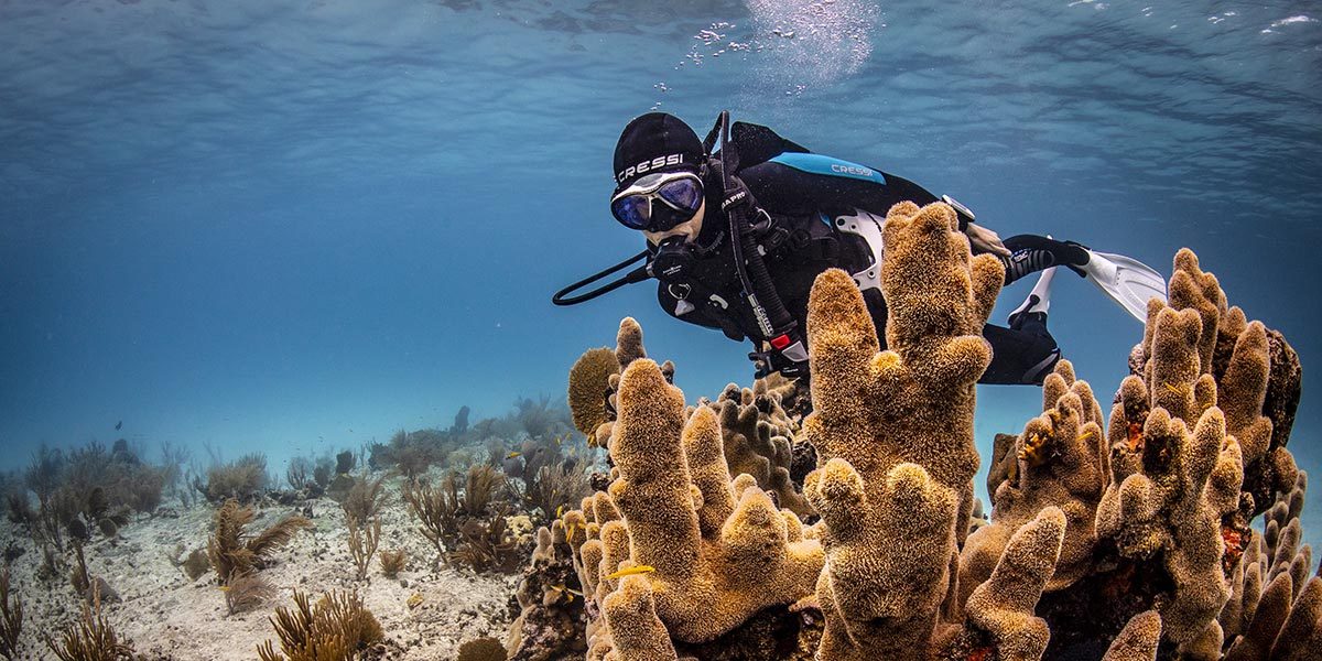
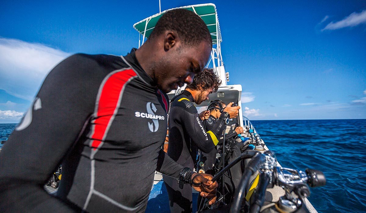
Cuts and scrapes are the most common injuries incurred by divers and snorkelers. DAN receives about one inquiry a week related to someone who has come into contact with coral. A burning sensation, pain and itching are common and may also be accompanied by a rash. These injuries can have a latent evolution and take weeks or months to heal, confusing both patients and clinicians.

Mechanisms of Injury

Soft living tissues cover the surface of corals. In the case of stony corals, the rigid (abrasive) structure underneath makes the coral’s soft tissue easy to tear and get into the scrape or cut. Foreign material can prolong the wound-healing process since the different antigens and substances cause an acute inflammatory process and infection. Cuts and scrapes from sharp-edged coral and barnacles tend to fester and may take weeks or even months to heal. Granulomas can form if debris from the original wound remains in the tissue. The body attempts to remove it, resulting in an itchy rash or papule (small, raised, tender bump) that lasts for some time before the body eliminates it.

While most “raspberries” generally heal quickly, skin abrasions from a marine environment can sometimes be more challenging to treat than those we get from outdoor activities such as baseball or bicycling. Whether it is a coral, a rock or a wreck, they all share a common factor: They are covered by living marine organisms, which makes coral cuts and scrapes unique.

Manifestations

The extent of the reaction depends on the presence and amount of toxins, the size and location of the abrasion and the pre-existing sensitivity of the injured person. The most common manifestations are a burning sensation, pain and itching. A rash may accompany the injury if the coral is a hydroid, such as fire coral.

Most animals of class Hydrozoa become hydroids as a life stage. They are predominantly colonial, and while most of them are marine creatures, you can find a few species in freshwater environments.

Fire corals are cnidarians, so they contain nematocysts. Touching them with a simple rub can cause mechanical activation and envenomation. The manifestation is usually blistering, which typically appears a few hours after contact. They typically resolve in a few days, but it is quite common for these injuries to relapse within a week or two after what seemed to be healing progress. This delayed reaction is typical of these types of envenomations.

Prevention

When underwater, try to avoid contact with coral or any other living creature. Whenever possible, wear a wetsuit or dive skin to protect yourself if you are accidentally pushed into coral by another diver or a current. Ocean divers should consider a marine animal first aid kit for their travels. Ready supplies will speed up the time to properly administer first aid for injuries. Additionally, for divers who want to learn more about the various marine life injuries, there are courses in marine life identification, first aid courses and a variety of books and publications available.

First Aid

  • Scrub the cut vigorously with soap and water, and then flush the wound with large amounts of water.
  • Flush the wound with a half-strength solution of hydrogen peroxide in water. Rinse again with water.
  • Apply a thin layer of antiseptic ointment, and cover the wound with a dry, sterile and non-adherent dressing. If you have no ointment or dressing, you can leave the wound open.
  • Clean and re-dress the wound twice a day.
  • If the wound develops a crust, use wet-to-dry dressing changes. Put a dry sterile gauze pad over the wound and soak it with saline or a diluted antiseptic solution (such as 1% to 5% povidone-iodine in disinfected water). Allow it to dry then rip the bandage off the wound. The dead and dying tissue should adhere to the gauze and lift free. The tissue underneath should be pink and may bleed slightly but should be healing. Change the dressings once or twice a day. Use wet-to-dry dressings for a few days or until they become non-adherent. Then resume the regular wound dressing described above.
  • Look for any signs of infection: extreme redness, red streaks on the extremity, pain, fever, pus or swollen lymph glands. If you have any, consult a qualified health professional about starting an antibiotic. A possible Vibrio bacteria infection can cause illness and even death in someone with an impaired immune system (e.g., from AIDS, diabetes or chronic liver disease).
  • Watch for coral poisoning, which can occur if abrasions or cuts are extensive or from a particularly toxic species. Symptoms include a wound that heals poorly or continues to drain pus, swelling around the cut, swollen lymph glands, fever, chills and fatigue. If you have these symptoms, see a physician.

Complications

The most frequent complications from non-stinging coral scrapes are inflammation (which leads to poor healing) and less commonly a secondary infection. Proper wound cleaning is crucial. If fire coral is the culprit, then a diluted acetic acid solution, such as household white vinegar, is a reasonable topical decontaminant and should be used as a soak to reduce the pain. Immersion in hot water can reduce the symptoms. Hot water is ideal, but you can use instant hot packs, cold packs or ice packs. Provide symptomatic treatment for the inflammatory response. Steroid creams are rarely helpful, and they can prolong a skin infection. If the inflammation is severe, you may administer systemic steroids in a moderate, tapering dose under the supervision of a trained medical provider. Oral antihistamines can sometimes help reduce the itching or burning sensation. 

Possible Complications of an Old Problematic Wound

It is not uncommon for divers to contact DAN concerned about a minor skin abrasion on their hands, knees or elbows that happened months ago and has not healed despite proper care. These chronic wounds often have a red and bumpy appearance, occasionally develop a crust and are usually painless. If common antibiotic ointments do not help, divers wonder if the cause may be a marine-specific pathogen.

Divers with an open wound, even a small cut or scrape, are at risk for skin infections. When an old problematic wound fits the descriptions above, it might have become infected with an opportunistic pathogen known as Mycobacterium marinum. Despite the name there are no marine-specific pathogens that affect humans. Some infections are more common in aquatic environments. M. marinum is responsible for a condition commonly known as fish tank granuloma, or aquarium granuloma.

The red and bumpy nodules, no larger than a centimeter, are granulomas — inflammatory immune cells trying to wall off the pathogen. Granulomas are usually isolated but can sometimes appear in small clusters. They are not necessarily painful. There may or may not be discharge from the wound.

Characteristics of M. marinum That Affect Healing

  • The pathogen is opportunistic. It causes infection only in the right conditions (environmental and patient-related), so it is often not considered as a potential culprit.
  • It likes cooler temperatures, which is why these wounds tend to flourish in areas with lower body temperatures such as hands, knuckles, elbows and knees.
  • Only specific antibiotics work, so the typical antibiotic treatments are usually unsuccessful.
  • The life cycle is slow, which means treatments last a long time. Sometimes patients will abandon what could have been a successful treatment or doctors may look for other potential explanations for the symptoms.
  • It requires specific culture media that a doctor would not ask for unless they suspected this pathogen. Standard culture results are often negative, which delays the diagnosis.

Allow your doctor to examine the wound and follow their standard procedures. The doctor will probably ask you how it happened or when it started. Tell them about the superficial abrasion in a marine environment. You may want to ask specifically if M. marinum could be the cause. Your doctor does not need dive-specific medical knowledge for this type of issue.

Fitness to Dive

Always take care of wounds and clean them thoroughly no matter the severity. The skin is our most effective and efficient means of immunological defense. A compromised wound can get seriously infected.

As a rule, treat wounds properly and let them heal before diving. This is particularly important before traveling to a remote location or one with limited local medical care capabilities. A skin lesion with the potential for infection might warrant a more conservative decision to stay ashore if you have such a trip planned.

Chronic skin lesions require specific consultation with your physician team before diving. Your doctor may prescribe treatment or a protective covering to prevent skin breakdown.

Sinus Barotrauma

Sinus barotraumas are among the most common diving injuries. When the paranasal sinuses fail to equalize to barometric changes during vertical travel, damage to the sinus can cause sharp facial pain with postnasal drip or a nosebleed after surfacing. Although sinus barotrauma is a prevalent and generally benign diving injury, some of its complications could pose a significant risk to the diver’s health. Divers should never underestimate difficulties equalizing sinuses.

Anatomy and Functions of the Paranasal Sinuses

The paranasal sinuses are gas-filled cavities in your facial bones and skull. They have several functions: They lighten the weight of your head, play a significant role in the resonance of your voice, serve as collapsible structures that protect vital organs during facial trauma, and may help the turbinates (small structures inside the nose) humidify and heat the air we breathe. There are two sets of four sinus cavities, one set on the right and one on the left.

  • The frontal sinuses (area one) are located within the forehead above your nose and eyes and are surrounded by thick, bony walls.
  • The ethmoid cells (area two) are located within the ethmoid bone between your eyes and nose and are formed by a variable number of connected individual cells.
  • The sphenoidal sinuses (area three) are centrally located behind the nasal cavity and vary in size and shape.
  • The maxillary sinuses (area four) are located within the maxillary bone below your eyes and lateral to your nose and are the largest pair of paranasal sinuses.
Paranasal sinuses. (Illustration by Michał Komorniczak)

The paranasal sinuses communicate with the nasal cavity via small orifices called ostia (singular: ostium). The ostia can easily be blocked by inflammatory processes, like colds or allergies, and in divers by improper attempts at equalization. Ostia blockage can impair drainage and make both descents and ascents troublesome.

Mechanisms of Injury

Every foot of descent in water adds approximately one-half pound of pressure on each square inch of tissue. The pressure diminishes by the same amount on ascent. According to Boyle’s Law, as the ambient pressure increases while descending, the volume of the gas in an enclosed space decreases proportionately. As the ambient pressure decreases while ascending, the volume of the gas increases proportionately.

While descending, it is imperative that divers actively or passively equalize all enclosed air-filled spaces to avoid injury. While ascending, the increasing volume usually vents itself passively. 

The mechanisms of injury of sinus barotraumas depend on whether it happened during descent or ascent.

During Descent (Squeeze)

Failure to equalize pressures on paranasal sinuses while descending keeps these cavities at atmospheric pressure, which results in a relative negative pressure (vacuum) as you descend to depth. The first sign of this type of sinus barotrauma is generally a sharp pain. The capillary vessels of the mucous membranes lining the sinuses engorge and burst, likely filling the sinuses with blood until the negative pressure is equalized. At this point the pain usually resolves or diminishes, and the diver continues the dive. While ascending, any remaining gas within the sinus expands and forces out this blood and mucus. These barotraumas usually manifest as postnasal drip or bloody discharge from the nose, depending on the sinuses involved. The bleeding can increase if you are taking blood thinners that include aspirin or other nonsteroidal anti-inflammatory drugs (NSAIDs).

During Ascent (Reverse Block)

Sinus barotrauma can also happen during ascent, known as a reverse block. Equalization of ears and sinuses during ascent is usually a passive event, which means active attempts should not be necessary. However, mild swelling and inflammation of the mucous membranes (as caused by a cold or by seasonal allergies) can compromise the narrow passages through which air escapes, trapping gas, mucus and blood. If a sinus fails to vent during ascent, the increasing pressure can apply significant tension to the mucosal lining and bony walls of the sinus. As the diver continues to ascend, one of the sinus walls can burst into an adjacent sinus that did vent correctly (the point of least resistance), effectively relieving the excess pressure. This type of sinus barotrauma manifests as a sharp facial pain during ascent, followed by a nosebleed or postnasal drip depending upon the sinus cavities involved.

Manifestations

The most common manifestations of sinus barotrauma are sharp facial pain during descent or ascent and blood dripping from the nose after surfacing. It is not uncommon for sinus barotrauma to be painless and manifest only as bloody mucus in the mask or the back of the throat.

Signs and Symptoms

Pain

  • Pain is usually facial in the region corresponding to the compromised sinus. In most cases the pain has a direct relation with changes in pressure on descent or ascent. In some cases the pain is delayed for a few hours; for example, when a sinus remains slightly over-pressurized following a dive.
  • Sharp pain in your forehead above and between the eyebrows is often a sign of barotrauma to your frontal sinuses. It is often described as an “ice-cream headache.” This type of sinus barotrauma usually has a direct relationship with changes in depth.
  • Pain behind your eyes is usually the result of a compromise to the ethmoidal sinus. You may also experience sharp pain, associated with changes in depth, behind and above the eyes.
  • Sharp pain beside your nose and below your eyes (upper maxillary region) is often a sign of maxillary sinus barotrauma. With changes in depth the pain might radiate to the upper molars or gums on the same side as the facial pain. The maxillary sinus and the upper jaw are supplied by the same nerve (trigeminal nerve).
  • Pain in the back (occipital region) or top of the head is the most intriguing, as its connection with the deeper sphenoidal cells is not obvious. When compared to the other sinuses, pain in the occipital region is often duller, like a normal headache. The association with changes in depth should be a clue that leads to a sinus origin.

Bleeding

  • You may notice some blood mixed with mucus and saliva in your mask after surfacing. You might not have been aware of it while diving. Minor bleeding that drips from the nose (technically not a nosebleed) or from the nose to the throat is typical of sinus barotrauma.
  • Minor bleeding is seldom a severe problem, but if you take an anticoagulant medication, be cautious when diving in a remote location. Uncontrolled bleeding without timely access to a medical facility prepared for such emergencies could be a severe health threat.

Coughing or Spitting Up Blood

While a nosebleed is not usually a manifestation of a life-threatening condition, postnasal drip usually results in blood in the diver’s mouth. This might be disconcerting to divers as it could be interpreted as the diver coughing up or spitting up blood. While there may be clues to determine whether this bloody discharge is of pulmonary origin or the result of sinus barotrauma, it is beyond the scope of what someone without medical training should attempt to evaluate. When in doubt, seek medical evaluation immediately.

Prevention

  • Do not dive when congested.
  • Refrain from diving when feeling fullness, pressure or pain in your paranasal sinuses.
  • Learn and use proper equalization techniques.

Medications

Talk to your doctor if you feel you need medication to dive. An ENT doctor is ideal for both ear and sinus problems, but your primary care physician can help with common problems. Using nasal sprays containing antihistamines and decongestants before diving may reduce swelling in the nasal and ear passages. Some are prescription only, while some are over the counter (OTC). With either option, your doctor may have special instructions on how to use them while diving.

Antihistamines prevent the effects of histamine, a substance produced and released by your body during the inflammations that cause nasal congestion, swelling of the mucous lining, and sneezing. While some of these drugs may cause drowsiness, second-generation antihistamines like cetirizine, loratadine and fexofenadine do not.

Decongestants relieve symptoms caused by the already-released histamine, clearing nasal and sinus congestion. Decongestants are not suitable for use by everyone. Some cardiovascular and central nervous system side effects could be concerns while diving.

Most nasal sprays work best if used one to two hours before the descent. They last from eight to 12 hours, so there is no need to take a second dose before a repetitive dive. Take short-acting nasal sprays like oxymetazoline 30 minutes before the descent; these usually last for 12 hours. Repeated use of short-acting OTC sprays can result in a rebound reaction that may set the stage for a reverse block. Steroid nasal sprays do not have this rebound effect but are slow-acting drugs, so you need to start them about a week in advance and use them regularly.

Whether you have a prescription or not, always check with your doctor before attempting to treat any condition.

Risk Factors

If you have a history of sinus trouble, allergies, a broken nose or deviated septum, or you currently have a cold, you may find the clearing procedure challenging to accomplish and may experience a problem with nosebleeds. It’s always best not to dive with a cold or any condition that may block the sinus air passages. If you experience difficulties during descent, this is the time to abort the dive. Remember that you can only abort a descent, never an ascent.

A good way to assess whether your paranasal sinuses are clear is by paying attention to your voice. You will sound like you have a stuffy nose due to a lack of appropriate nasal airflow while speaking.

Being able to breathe through your nose only proves your nasal passages are clear. It does not indicate anything about your paranasal sinuses.

Complications

With this type of injury, blood can run down the back of the throat or pool in the sinuses below the eyes and emerge later (even days after diving) as a thick, black, bloody discharge. The collected blood can also act as a growth medium for bacteria and result in sinus infections. 

Pneumocephalus (air between the skull and the brain) and orbital emphysema (air behind and around the eyeball) are rare but important complications of sinus barotraumas. If not adequately treated, they may cause serious neurological and life-threatening complications. Never underestimate sinus barotrauma. 

First Aid

  • Use a nasal decongestant spray or drops. This might reduce the swelling of the mucous membranes, which may help to open the ostia and drain fluid from the sinuses.
  • Seek professional medical evaluation. Any doctor should be able to help, regardless of any dive medicine knowledge or training.

Implications in Diving

For the Diver

  • You can consider a return to diving if a physician determines that the injury has healed, and the risk of further injury is no greater than normal.
  • Do not neglect these injuries. Some of the complications could negatively affect you for the rest of your life.

For the Dive Operator

  • Provide first aid treatment, as described above. As the expedition’s leader, you have a duty of care for a diver injured during your trip.
  • Be skeptical of any folkloric first aid treatments. Use common sense, and don’t attempt any magic solutions. Remember that you might be liable.
  • Have them evaluated by a medical professional in a timely fashion.
  • Don’t worry about referring them to a doctor with dive medicine experience. An ENT specialist is ideal, but any doctor should be able to help.
  • Do not allow any further diving once the injury has occurred until they are cleared by a physician.

For the Physician

  • Provide symptomatic treatment (anti-inflammatory drugs, decongestants, mucolytic agents).
  • Prophylactic antibiotic therapy is controversial. Although a middle-ear infection is a plausible secondary complication, this is not always the case in the acute phase.
  • Assess concomitant middle-ear barotrauma.
    • If present, consider referring the patient to an ENT specialist.
    • Use the O’Neill grading system or detail what you observe.
  • Assess the cranial nerve function.

Fitness to Dive

Do not dive until swelling and inflammation have resolved, and you can adequately equalize, preferably under otoscopic evaluation. Assess why the problem occurred (lack of training, allergy, etc.) and address each factor. The inability to equalize properly is disqualifying.

If you are unable to clear your sinuses or you have frequent nosebleeds when diving, you should see your primary care physician or an ear, nose and throat (ENT) specialist (otolaryngologist) for evaluation.

Juggling Physical Exercise and Diving

exercising with kettle bells

If you want to dive, you need to be ready. Readiness entails medical, psychological and physical fitness, appropriate knowledge and adequate physical skills. If you exercise regularly at an intensity that keeps your heart rate above 70 percent of maximum or so for more than 90 minutes per week, it is a good bet that you are physically fit enough to dive recreationally under a variety of conditions. Just diving, however, will likely not be enough to constitute regular exercise. In addition, exercise conducted during or close to diving has implications for safety. By following the right recommendations and protocols, you can ensure strength and safety for diving all year long.

Physical Fitness for Diving

Divers need to have sufficient strength and aerobic capacity reserves to meet both the normal and reasonable exceptional demands of diving in their chosen environment.7 Physical fitness is maintained when the intensity and frequency of exercise are sufficient to protect the body’s capacity — the array of biochemical and physiological capacities that determine a fitness limit. Physical fitness is improved when the exercise load exceeds the body’s current capacity and a training effect is established. Most training programs rely on progressive overload — the incremental increase in training intensity to continue the drive to adapt at a pace that can be tolerated. Exceeding the threshold for maintenance or improvement of fitness, as desired, makes for an effective workout.

While a diver’s physical strength can be tested by carrying tanks and related gear, the duration of the effort is typically too short to constitute an effective workout. The aerobic demands of most well-planned dives are even less likely to reach the intensity to protect even a moderate aerobic capacity. Ultimately, the diver has to do something outside of normal diving to maintain or improve their fitness level.

There are additional fitness issues directly relevant to diving physiology. While the data are incomplete, physical fitness has been associated with fewer post-decompression bubbles in humans.2,8 While bubbles are not equated with decompression sickness, it is accepted that lower bubble counts indicate a reduced degree of decompression stress. Experiencing less decompression stress on any dive is definitely a good thing. Animal models have also demonstrated a lower incidence and reduced severity of decompression sickness (DCS) for trained versus untrained subjects.1,10 Ultimately, it may be clear that sound physical fitness is desirable for decompression safety as well as for physical competence, which is the ability to meet the physical demands of a situation.

Timing Exercise and Diving

Just as important as a regular exercise regimen is the timing of such as it relates to diving. Scheduling outside physical fitness activities can be problematic when someone dives frequently. While part of this is simply a time management problem, there are other considerations. Conducting intense physical exercise too close to diving activity can be problematic for more fundamental reasons.

Bubble formation, while noted earlier as not equivalent to or a guarantee of DCS, can indicate an increased risk for it. Intense physical activity — generally with substantial muscular forces and joint loading, or the application of forces on joints — is believed to transiently increase micronuclei activity, the presumed agent of bubble formation. Intense physical activity too close to diving may therefore be problematic. Physical activity after diving may also stimulate additional bubble formation, possibly through a combination of increased microicronuclei activity and increased joint forces.

Interestingly, some preliminary work has shown that an intense bout of exercise conducted 24 hours prior to diving may reduce bubble presence in humans,3 possibly by inhibiting micronuclei activity. This potentially protective effect was not seen with exercise conducted closer to dive time. While this effect needs to be validated, the preliminary findings may support a simple rule of thumb for scheduling exercise. To reduce the risk, it is a good idea to avoid intense exercise 24 hours before and after diving.

The near-dive window will be best for low-intensity activities. Those who participate in cross-training activities may find it easiest to accommodate this schedule. For those who are more singleminded, diving may fit well into training rest days for those who put the priority on exercise, while training may fit best into diving rest days for those who put the priority on diving. Overall, lower training intensities will likely be more appropriate for the latter group, but accommodations can be reached.

Timing of Exercise During Diving

Physical activity during the dive also has a direct impact on decompression safety.4,5,6,9 Exercise during the compression and bottom phase increases inert gas uptake, effectively increasing the subsequent decompression obligation of any exposure. It is important to remember that dive tables and computers estimate inert gas uptake, they never know reality. However, light exercise during the decompression phase (including safety or decompression stops) increases inert gas elimination and reduces risk. The caveat regarding exercise during decompression is that more is not always better. Too much or too intense exercise during the decompression phase can stimulate bubble formation, thus inhibiting inert gas elimination and increasing decompression risk.

Final Recommendations

We do not yet have sufficient data to quantify the difference between beneficial and potentially harmful exercise. Understanding the various issues and applying common sense offer the best protection. Most important is that moderate time-depth profiles are your best defense. Exercise considerations provide only a secondary defense. In terms of the secondary defense, though, the compression and bottom phases are best associated with the lightest exercise possible. Ascent and stop phases are best associated with mild, low-intensity exercise. Exercise that is aggressive and/or stimulates substantial joint-loading is almost always undesirable at any point near or during a dive.

The post-dive period is a good time to take it easy. Both decompression safety and mental health can be helped by an extended period of relaxation between the end of the dive and the start of equipment shifting and/or racing on to the next activity.

Physical fitness — including both strength and aerobic capacity — is important for divers both for physical safety and decompression safety. Regular exercise training is best scheduled to separate intense exercise and diving. Intense physical training should be avoided 24 hours on either side of diving activity. Any exercise within 24 hours of diving should involve the lowest possible joint forces.

Neal Pollock, Ph.D.

References

1. Broome JR, McNamee GA, Dutka AJ. “Physical conditioning reduces the incidence of neurological DCI in pigs.” Undersea Hyperb Med. 1994; 21(suppl): 69.

2. Carturan D, Boussuges A, Burnet H, Fondarai J, Gardette B. “Circulating venous bubbles in recreational diving: relationships with age, weight, maximal oxygen uptake and body fat percentage.” Int J Sports Med. 1999; 20(6): 410-414.

3. Dujic Z, Duplancic D, Marinovic-Terzic I, Bakovic D, Ivancev V, Valic Z, Eterovic D, Petri NM, Wisloff U, Brubakk AO. “Aerobic exercise before diving reduces venous gas bubble formation in humans.” J Physiol. 2004; 555(3): 637-642.

4. Jankowski LW, Nishi RY, Eaton DJ, Griffin AP. “Exercise during decompression reduces the amount of venous gas emboli.” Undersea Hyperb Med. 1997; 24(2): 59-65.

5. Jankowski LW, Tikuisis P, Nishi RY. “Exercise effects during diving and decompression on postdive venous gas emboli.” Aviat Space Environ Med. 2004; 75(6): 489-495.

6. Jauchem JR. “Effects of exercise on the incidence of decompression sickness: a review of pertinent literature and current concepts.” Int Arch Occup Environ Health. 1988; 60(5): 313-319.

7. Pollock NW. “Aerobic fitness and underwater diving.” Diving Hyperb Med. 2007; 37(3): 118-124.

8. Powell MR. “Exercise and physical fitness decrease gas phase formation during hypobaric decompression.” Undersea Biomed Res. 1991; 18(suppl): 61.

9. Van der Aue OE, Kellar RJ, Brinton ES. “The effect of exercise during decompression from increased barometric pressures on the incidence of decompression sickness in man.” US Navy Experimental Diving Unit Research Report No. 8-49, 1949.

10. Wisloff U, Brubakk AO. “Aerobic endurance training reduces bubble formation and increases survival in rat exposed to hyperbaric pressure.”

Swimmer’s Ear (Otitis Externa)

divers preparing to dive

Swimmer’s ear (otitis externa) is a condition caused by inflammation or infection of the outer ear canal. In a diving environment, this is usually caused when prolonged exposure to wet conditions changes the natural acidity and flora in the ear canal, allowing opportunistic bacteria or fungi to grow and become pathogens. Warm and humid environments and excessive cleaning of the ear canal can predispose a person to otitis externa.

Epidemiology

  • Swimmer’s ear affects one in 200 Americans every year and is a chronic problem in 3 to 5 percent of the population.
  • Swimmers, surfers and other people who are exposed to wet and warm conditions are at increased risk.

Symptoms

The main symptoms are ear pain, warmth and itching — often in the ear canal . Occasionally, the pinna (the cartilaginous external part of the ear) may show signs of inflammation with redness, swelling and pain. If left untreated, swelling can increase to include nearby lymph nodes and produce enough pain that movement of the jaw may become uncomfortable or painful.

  • Pain, warmth and itching in the ear canal
    • Pain when moving the jaw or when gently pulling on the earlobe or pushing on the pinna
  • Muffled hearing (transient, usually a sign of concomitant middle ear compromise)
  • Discharge from the ear canal is possible if initial symptoms are neglected for a few days

Management & First Aid

Swimmer’s ear is often self-diagnosed. Professional medical diagnosis is often clinical as well, not requiring more than an ear exam with an otoscope. With proper treatment, symptoms often resolve within a few days, rarely taking longer than a week.

  • Do not neglect the initial itching or mild pain, as it may progress overnight.
  • Seek a professional medical evaluation. Although diagnosing swimmer’s ear is straightforward, proper treatment requires a prescription of otic antibiotic drops.
  • Once you have symptoms, do not introduce any preventative drops or preparations.

Prevention

Keep your ears clean and dry. Although it might be tempting to keep your ears clean using cotton swabs or similar, the best way to avoid problems with your ears is to not mess with them. Wash your ears with regular soap when you take a shower and don’t insert anything in them. Under normal circumstances, this is all we need.

Your ears produce a waxy substance that is hydrophobic to prevent moisture retention and acidic enough to prevent bacterial growth. Excessive moisture — as is common with frequent diving — can cause an emulsification of the natural ear wax, which can change the environment in the ear canal to make it more susceptible to infections.

  • Dry your ears with a towel after swimming, showering or diving. Tilting your head and pulling your earlobe in different directions while your ear is facing down might help eliminate water.
  • A hair dryer could be used to carefully dry the ear after a shower. Be careful to ensure the air is not too hot, hold it at least a foot (about 30 centimeters) away from the ear.
  • Refrain from putting objects (such as cotton swabs or ear wax removal tools) in the ear canal. This can cause ear wax (cerumen) impaction and can damage the skin in the ear, potentially increasing the risk of infection.
  • Excessive debris or cerumen may trap water in the canal.
    • If you think you have an excess of buildup ear wax, consult your doctor and allow a medical professional to do a proper cleaning. Remember that cleaner is not necessarily better. Washing the ear canal excessively will change the acidity of the ear canal, and a less acidic environment is a common cause of opportunistic pathogen growth that could lead to an ear infection.
  • Talk to your doctor about whether you should use any alcohol-based ear drops after swimming or diving.

Prognosis & Returning to Dive

Prognosis is generally good if the condition is addressed promptly. Upon determination by your physician that the infection has resolved, you may return to diving.

WARNING: Continuing to dive with unresolved swimmer’s ear will perpetuate the condition that caused an infection in the first place, allowing pathogens to prolong and worsen the infection.

Inner-Ear Barotrauma (IEBT)

Inner-ear barotrauma is damage to the inner ear due to pressure differences, usually caused by incomplete or forceful equalization. A leak of inner-ear fluid (perilymph fistula) may or may not occur.

Blausen.com staff (2014). “Medical gallery of Blausen Medical 2014”. WikiJournal of Medicine 1.

Anatomy and Functions of the Ear

The human ear has three distinct sections:

  • External Ear
    This includes the ear itself and the ear canal to the eardrum.

  • Middle Ear
    This is an air-filled cavity between the eardrum and the inner ear. It has three components: the middle-ear cavity, the three ear bones (ossicles) and the mastoid process.

  • Inner Ear
    The inner ear is a sensory organ. It is part of the central nervous system, and it has two functions:

    • Auditory: The cochlea turns soundwaves into electrical impulses for the brain.
    • Balance, orientation and acceleration: The canals provide some of our control of balance and position and help detect acceleration.

Mechanisms of Injury

When you properly equalize the pressure in your middle ear, your risk of inner-ear barotrauma is very low. If you do not equalize the pressure in the middle ear during descent, so the water pressure on the eardrum transfers inward and may damage sensitive inner-ear structures. If the pressure is excessive, the oval window or, more commonly, the round window may tear, and the inner-ear fluid may leak into the middle ear. This is known as a perilymph fistula.

The Valsalva maneuver is a common equalization technique. This maneuver increases the intrathoracic pressure, which means the pressure is exerted on all intrathoracic organs. Pliable blood vessels such as the superior vena cava transmit this increased pressure into the head. The skull is a rigid structure with no capacity to expand, so the result is an increase in intracranial pressure. The cochlea is a fluid-filled organ surrounded by soft tissues as well as bone; its only weak point is the external wall in the vestibule, which is adjacent to the middle-ear space. The round and oval windows are two thin and delicate tissues that reverberate with sound. As the increased pressure transmits through the cochlear fluid, it causes an outward movement of the round window.

Pressure waves alone can cause damage to the inner ear without window rupture. If a rupture occurs, the loss of fluid from the inner ear leads to damage to the vestibular system, causing sudden hearing loss and often acute vertigo with loss of balance. If the leak is not stopped soon by spontaneous healing or surgical repair, permanent hearing loss may occur.

Manifestations

Symptom onset is usually sudden and often associated with ear equalization issues. Symptoms of middle-ear barotrauma are often present, but their absence does not rule out inner-ear barotrauma. Vertigo is usually severe and accompanied by nausea and vomiting. Hearing loss can be complete, instant and permanent. Hearing loss in divers usually manifests as loss of higher frequencies (acute, high-pitched sounds). The loss might become noticeable only after a few hours. Divers may not be aware of the loss until they have a hearing test.

Signs and Symptoms

  • Ear pain may or may not be present.
  • A severe onset of vertigo (spinning sensation) is usually present.
  • Loss of spatial orientation is possible.
  • Hearing loss, sometimes with tinnitus (ringing in the ears), may occur.
  • The eyes might show nystagmus (involuntary rapid and repetitive eye movement).
  • A feeling of fullness in the ears (often the least of the diver’s complaints) is possible.

Prevention

  • Do not dive when congested.
  • Refrain from diving when feeling popping or crackling in your ears, or if you have a feeling of fullness in your ears after diving.
  • Learn and use proper equalization techniques.

First Aid

  • If you have symptoms of inner-ear barotrauma, do not try to equalize your ears (even if you feel fullness in them, which is likely). This might make things worse.
  • Use a nasal decongestant spray or drops. This might reduce the swelling of the mucous membranes, which may help to open the Eustachian tubes and drain the fluid from the middle ear.
  • Do not put any drops in your ear canal. If the tympanic membrane is intact, the drops will do nothing. If the tympanic membrane is ruptured, drops might make things worse.
  • Lying down and closing your eyes may help with vertigo, which might be significant and will likely make you feel miserable. Try to remain calm. Vertigo is usually accompanied by nausea and vomiting.
  • First aid providers should administer oxygen.
  • Seek professional medical evaluation ASAP. Any doctor should be able to help, regardless of any dive medicine knowledge or training.
  • First aid providers should conduct a complete neurological exam and note any deficits. It is important to differentiate inner-ear barotrauma from inner-ear decompression sickness if possible.
  • An ear, nose and throat (ENT) specialist (otolaryngologist) might be the most qualified physician to help in this situation.

Implications in Diving

For the Diver

  • Follow the first aid recommendations above.
  • Avoid rapid head movements.
  • Avoid any exertion, middle-ear equalization, diving, altitude exposure, sneezing and nose-blowing.
  • Do not lift heavy weights; Valsalva-like maneuvers might exacerbate vertigo.
  • Lie down and rest. Keep movement and physical activity to a minimum.
    • Vertigo might be accompanied by nausea and vomiting. Lie on your side to avoid aspirating vomit.
  • It may take time to return to diving, and you should resume diving only after proper evaluation from a physician with experience in diving medicine, usually in consultation with an ENT specialist.
  • Do not neglect these injuries. Some possible complications may have a negative impact on normal living.

For the Dive Operator

  • Provide first aid treatment, as described above. As the expedition’s leader, you have a duty of care for a diver injured during your trip.
    • Be skeptical of folkloric first aid treatments. Use common sense, and don’t attempt magic solutions. Remember that you might be liable.
  • Have the diver sit down, and reassure them during the process.
  • Help them deal with vertigo, which can be a very uncomfortable feeling that will likely make the diver — and you — feel uneasy about the situation. Rapid movements of the head and Valsalva-like maneuvers (such as lifting heavy things) might exacerbate vertigo. People with vertigo usually have:
    • A spinning sensation: They feel they are spinning or that the environment is spinning around them.
    • Repetitive nystagmus: Involuntary eye movement that can occur from side to side, up and down, or in a circular motion.
    • Nausea and vomiting: Make sure the diver does not aspirate vomit.
  • Have the diver evaluated by a medical professional in a timely fashion.
  • Don’t worry about finding a doctor with dive medicine experience. An ENT specialist would be ideal, but any doctor should be able to help with the initial evaluation.

For the Physician

  • Assess for middle-ear barotrauma.
  • Differentiate between IEBT and IEDCS.
  • Assess vestibular function.
    • Vertigo, nystagmus and/or hearing loss might be suggestive of IEBT.
  • Assess the eighth cranial nerve.
  • Strongly discourage your patient from continuing to dive until evaluated by a specialist.
  • Consider conservative treatment, including bed rest in a sitting position and avoiding any straining that nay increase intracranial or middle-ear pressure.
  • Discourage physical exertion, including lifting heavy things.
    • Valsalva-like maneuvers might induce more vertigo if a perilymphatic fistula is present.

IEBT or Inner-Ear Decompression Sickness (IEDCS)?

It is important to distinguish between these two conditions because their treatments differ. The standard treatment for DCS of any kind is hyperbaric oxygen therapy in a recompression chamber. Recompression (or any pressure change) is contraindicated when inner-ear barotrauma is likely. Differential diagnosis between IEDCS and IEBT can sometimes be a challenge. While the symptoms are similar in both conditions, there are a few characteristics that might help during the assessment.

IEBT

  • Often preceded by failed equalization of middle-ear pressure
  • Usually very acute symptom onset (immediate to a few minutes)
  • Usually at the beginning of a dive (during descent) as a result of difficulty equalizing
  • Evidence of middle-ear barotrauma — check the tympanic membranes

IEDCS

  • Often more delayed symptom onset (many minutes to a few hours)
  • Usually the result of a failed decompression after a moderate to significant dive exposure
  • Can be associated with a patent foramen ovale (PFO)
  • Other forms of DCS, including cutaneous DCS, may be observed

Fitness to Dive

Do not dive until the injury is healed, and you can adequately equalize, preferably under otoscopic evaluation. Assess why the problem occurred (lack of training, allergy, etc.) and address each factor. If you are unable to equalize, then you may consider ENT consultation. The inability to equalize properly is disqualifying.

Note: Do not dive with earplugs, as this may cause external-ear barotrauma.

Cardiovascular Fitness and Diving

adults taking a cycling class

Scuba diving exposes you to many effects, including immersion, cold, hyperbaric gases, elevated breathing pressure, exercise and stress, as well as a post-dive risk of gas bubbles circulating in your blood. Your heart’s capacity to support an elevated blood output decreases with age and with disease. Having a healthy heart is of the utmost importance to your safety while scuba diving as well as to your ability to exercise generally and your life span.

Below are some common cardiovascular issues that could affect a diver, including their implications in diving and possible medical treatments.

Hypertension

Hypertension, or high blood pressure, is one of the most common medical conditions seen in the diving population — no surprise since it is a common medical condition in the general population. Strict criteria for hypertension can vary depending on the reference cited, but normal blood pressure is generally accepted to be a systolic pressure below 140 and a diastolic pressure below 90 mm Hg, depending on age (cited as systolic first and diastolic second — e.g. “120 over 80,” by your doctor). A thorough medical evaluation should be performed to find a treatable cause for hypertension; in most cases, however, none will be found.

Basically, two different sets of complications face a person with hypertension: short-term and long-term. Short-term complications are generally due to extremely high blood pressure; the most significant is the risk of a stroke due to rupture of blood vessels in the brain (called a cerebrovascular accident). Long-term detrimental effects are more common: they include coronary artery disease, kidney disease, congestive heart failure, eye problems and cerebrovascular disease.

Implications in Diving: 

As long as the individual’s blood pressure is under control, the main concerns should be the side effects of medication(s) and evidence of end-organ damage. Divers who have demonstrated adequate control of blood pressure with no significant decrease in performance in the water due to the side effects of drugs, should be able to dive safely.

A recent report in a diving medical journal citing several episodes of acute pulmonary edema (i.e., lungs congested with fluid) in individuals with uncontrolled hypertension while they were diving. Regular physical examinations and appropriate screening for the long-term consequences of hypertension such as coronary artery disease are necessary.

Medication Used in Treatment: 

Mild hypertension may be controlled with diet and exercise; however, medication is often necessary. Many classes of drugs are used to treat hypertension, with varying side effects. Some individuals must change medications after one drug appears to be or becomes ineffective. Others might require more than one drug taken at the same time to keep the blood pressure under control.

Classes of drugs known as beta-blockers often cause a decrease in maximum exercise tolerance and may also have some effect on the airways. This normally poses no problem for the average diver. ACE (angiotensin converting enzyme) inhibitors are the preferred class of drugs for treating hypertensive divers; a persistent cough is a possible side effect.

Calcium channel blockers are another choice, but lightheadedness when going from a sitting or supine position to standing may be a significant side effect.

Diuretics are also frequently used to treat hypertension. This requires careful attention to hydration and electrolyte status. Most anti-hypertensive medications are compatible with diving as long as the side effects experienced by the diver are minimal and their performance in the water is not significantly compromised. Any diver with long-standing high blood pressure should be monitored for secondary effects on the heart and kidneys.

Coronary Artery Disease

Coronary atherosclerosis is commonly described as “hardening of the arteries.” It’s the result of the deposition of cholesterol and other material along the walls of the arteries of the heart. The walls of the arteries, in response to the deposition of this material, also thicken. The end result is a progressively increasing blockage to blood flow through the vessel. Many factors contribute to the development of coronary atherosclerosis: a diet high in fat and cholesterol, smoking, hypertension, increasing age and family history. Women of reproductive age are generally at a lower risk due to the protective effects of estrogen. In the United States and other industrialized countries, coronary artery disease is the leading cause of death.

Implications in Diving: 

Symptomatic coronary artery disease is a contraindication to safe diving: don’t dive with it. Coronary artery disease results in a decreased delivery of blood — and therefore, oxygen — to the muscular tissue of the heart. Exercise increases the heart’s need for oxygen. Depriving myocardial tissue of oxygen can lead to abnormal heart rhythms and/or myocardial infarction, or heart attack.

The classic symptom of coronary artery disease is chest pain, especially when it follows exertion. Unfortunately, many people have no symptoms before they experience a heart attack. Cardiovascular disease is a significant cause of death among divers. Older divers and those with significant risk factors for coronary artery disease should have regular medical evaluations and appropriate studies (e.g., treadmill stress test).

Medication Used in Treatment: 

Medications typically used in the treatment of this disease include nitroglycerin, calcium channel blockers and beta-blockers. At some point, someone with coronary artery disease may need a revascularization procedure, or the re-establishment of blood supply, through bypass surgery or angioplasty. If the procedure is successful, the individual may be able to return to diving after a period of healing and a thorough cardiovascular evaluation. (See “Coronary Artery Bypass Grafting,” below)

Myocardial Infarction (Heart Attack)

Myocardial infarction (MI), or heart attack, occurs when damage to the heart muscle cells results from interrupted blood flow to the tissue. Risk factors for heart attack are the same as those for coronary artery disease.

Most commonly, a myocardial infarction is the direct consequence of coronary atherosclerosis, or hardening of the arteries. The blocked arteries stop blood flow to the heart tissue and deprive the cells of necessary oxygen. Small areas of heart muscle may sustain damage, resulting in a scar; this may even occur without the individual experiencing significant symptoms. If larger areas of the heart are deprived of oxygen or if the cells that conduct the primary electrical impulses are within an area where blood flow is decreased, the heart may beat irregularly or even stop beating altogether. It is not unusual for sudden cardiac death to be the first symptom of coronary artery disease.

Implications in Diving: 

Cardiovascular events cause 20 to 30 percent of all deaths that occur while scuba diving. For many people, the real problem is that the first sign of coronary artery disease is a heart attack. The only realistic approach is to recommend appropriate measures to prevent the development of coronary atherosclerosis and to encourage regular medical evaluations for those individuals at risk.

Prudent diet and regular exercise should be habitual for divers. Older individuals and divers who have a family history of myocardial infarctions, especially at an early age, should receive appropriate evaluations to detect early signs of coronary artery disease.

Individuals who have experienced previous heart attacks are at risk for additional cardiac events in the future, and damaged heart tissue may have compromised cardiac function. The damaged left ventricle may not be able to pump blood as efficiently as it could prior to the MI.

Regardless of whether an individual has had a revascularization procedure (see “Coronary Artery Bypass Grafting”), strict criteria must be met prior to an individual’s safe return to diving. After a period of healing — six to 12 months is recommended — an individual should undergo a thorough cardiovascular evaluation, which includes an exercise stress test. The individual should perform at a level of 13 mets (stage 4 on Bruce protocol). This is a fairly brisk level of exercise, equating to progressively running faster until the patient reaches a pace that is slightly faster than running an 8-minute mile (for a very brief period of time). Performance at that level without symptoms or EKG changes indicates normal exercise tolerance.

Coronary Artery Bypass Graft

Fortunately, for both patients and thoracic surgeons coronary artery disease affects the first part, or proximal end, of the artery much more frequently and severely than the downstream portion of the artery. This allows for a surgical procedure that uses a portion of a vein or another artery to direct blood around the blockage. Doctors perform this procedure hundreds of times daily around the country – more than 500,000 times annually. If the bypass is successful, the individual should become free of the symptoms of coronary artery disease, and the heart muscle should receive normal blood flow and oxygen.

A less invasive procedure, coronary angioplasty, consists of placing a catheter with a balloon on its tip into the area of the blockage and inflating the balloon to open the artery. This procedure does not require opening the chest and can be performed in an outpatient setting.

Implications in Diving: 

An individual who has undergone coronary artery bypass grafting or angioplasty may have suffered significant cardiac damage prior to having the surgery. The post-operative cardiac function of individuals dictates their fitness for diving.

Anyone who has had open-chest surgery needs appropriate medical evaluation prior to scuba diving. After a period of stabilization and healing (6-12 months is usually recommended), the individual should have a thorough cardiovascular evaluation prior to being cleared to dive. He or she should be free of chest pain and have normal exercise tolerance, as evidenced by a normal stress EKG test (13 mets or stage 4 of the Bruce protocol — defined at the end of previous section on MI). If there is any doubt about the success of the procedure or how open the coronary arteries are, the individual should refrain from diving.

Mitral Valve Prolapse

Mitral valve prolapse (MVP) is a common condition, especially in women. The problem arises from some excess tissue and loose connective tissue in the structure of the mitral valve in the heart: part of the valve protrudes down into the left ventricle during contraction of the heart.

An individual with MVP may have absolutely no symptoms, or the symptoms may vary from occasional palpitations, or unusual feeling in the chest arising from the heart beating, to atypical chest pain and a myocardial infarction. There is also a slightly increased risk of a small stroke or transient loss of consciousness.

Implications in Diving:

Frequently mitral valve prolapse will not cause any symptoms or result in any changes in blood flow that would prevent an individual from diving safely. A diver with known mitral valve prolapse who has no symptoms and takes no medications for the problem should be able to safely participate in diving. The individual should require no medications and should be free from chest pain, any alteration in consciousness, palpitations and abnormal heartbeats. Individuals with abnormal cardiac rhythm, which can produce palpitations, should not dive unless these palpitations can be controlled with low doses of anti-arrhythmic medications.

Medication Used in Treatment: 

Beta-blockers are occasionally prescribed for mitral valve prolapse. These often cause a decrease in maximum exercise tolerance and may also have some effect on the airways. This normally poses no problem for the average diver, but it may be important in emergency situations.

Dysrhythmias

The term “dysrhythmia” means abnormal heartbeat and is used to describe a wide range of conditions ranging from benign, non-pathologic conditions to severe, life-threatening rhythm disturbances. More familiar to many people is the term “arrhythmia,” which literally means “no heartbeat.”

The normal heart beats 60 to 100 times each minute. In well-trained athletes or even in select non-athletic individuals completely at rest, the heart may beat as slowly as 40 to 50 times each minute. Entirely healthy, normal individuals have occasional extra beats or minor changes in rhythm. These can be caused by drugs (caffeine), stress, or for no apparent reason. Dysrhythmias become serious only when they are prolonged or when they do not result in the desired mechanical contraction of the heart.

Physiologically significant extra heartbeats may originate in the upper chambers of the heart (supraventricular tachycardia or atrial dysrhythmia) or in the lower chambers of the heart (ventricular tachycardia). The cause may be due to a short-circuit or an extra conduction pathway for the impulse or secondary to some other cardiac pathology. People who have episodes or periods of rapid heartbeat are at risk for losing consciousness during these events. There are also conditions where the person has a fairly stable dysrhythmia (e.g., fixed atrial fibrillation), but they usually have additional cardiovascular and other health problems that coincide with their rhythm disturbance. A slow heart rate or heart block may cause symptoms, too.

Implications in Diving: 

The more serious dysrhythmias, like ventricular tachycardia and many types of atrial rhythm disturbances, are incompatible with diving. The risk for any person developing a dysrhythmia during a dive is, of course, losing consciousness while underwater. Supraventricular tachycardias are unpredictable in onset and are often triggered by immersing the face in cold water. Someone who has had more than one episode of this type of dysrhythmia should not dive.

An individual with any cardiac dysrhythmia needs a complete medical evaluation by a cardiologist prior to engaging in scuba diving. In some cases, thorough conduction (electrophysiologic) studies can identify an abnormal conduction pathway and the problem can be corrected. Recently, doctors and researchers have determined that people with some dysrhythmias (e.g., certain types of Wolff-Parkinson-White Syndrome) may safely participate in diving after a thorough evaluation by a cardiologist. Also, in select cases, some people with stable atrial dysrhythmias (e.g., uncomplicated atrial fibrillation) may dive safely if a cardiologist determines that there are no other significant health problems.

Medication Used in Treatment: 

Most dysrhythmias that require medication are medically disqualifying for safe diving. Exceptions may be made on a case-by-case basis in consultation with a cardiologist and diving medical officer.

Murmurs

A heart murmur is an extra sound that can be heard during chest examination with a stethoscope. The opening and closing of the heart valves produce expected and predictable sounds in individuals with normal heartbeats. Murmurs represent extra sounds caused by turbulent or abnormal flow of blood past a heart valve, in the heart itself or in great vessels (i.e., aorta, pulmonary arteries).

Some murmurs occur strictly from increased flow. For example, pregnant women often have a functional murmur due to a greater blood volume and hyperdynamic metabolism; these are benign. Other murmurs are due to damaged heart valves and represent significant pathology. Damaged valves may either restrict blood flow (stenotic lesions) or allow blood to flow back into the chamber of the heart from which it had just exited (regurgitant lesions). Heart valves can be damaged due to infection, trauma, heart muscle damage (myocardial infarction), or an individual may be born with a structurally abnormal heart valve.

Implications in Diving:

Stenotic lesions, such as aortic and mitral stenosis, restrict efficient blood flow and may have serious consequences during exercise. Significant aortic stenosis places an individual at greater risk for sudden cardiac death while exercising; it is a contraindication for diving. Mitral stenosis also limits the response to exercise and, over a period of time, can result in congestive heart failure.

Regurgitant lesions pose somewhat less of a risk during diving. Over a period of years, the heart will be taxed by the extra work necessary to pump blood, and heart failure may be the long-term result. Divers with these types of heart valve problems may safely participate in diving if they have no symptoms and have normal left ventricular structure and function, as evidenced by an echocardiogram.

Atrial And Ventricular Septal Defects

An atrial septal defect (ASD) results from the incomplete closing of the wall that separates the right and left atria (the two upper chambers of the heart) during embryonic development. This is not an uncommon phenomenon in the general population, and, if the hole is small enough, the average person will experience minimal physiologic consequences. Women are affected more commonly than men.

Surgical correction of the defect may be undertaken, especially if the person is experiencing symptoms secondary to blood flowing from the normally higher pressure left atrium to the right atrium. Early in life, symptoms may be few, but over a period of years, complications, such as abnormal heart beats and shunting (bypassing) of blood from left to right may occur.

On examination, the person with an ASD may have a significant murmur. A ventricular septal defect (VSD) is a communication, or opening, between the right and left ventricles, the lower chambers of the heart. A fairly common developmental abnormality, VSD often merits surgical correction if the defect is large. Because of the large difference in pressures between the left and right ventricles, blood flow through the defect is nearly always from left to right. The individual with ventricular septal defects may experience long-term consequences.

Implications in Diving: 

While the normal pressures in the chambers of the heart favor blood flowing from left to right through an ASD and VSD, periods in which this flow is reversed can occur, particularly for ASD. Although individual variations exist, Doppler studies have shown that most divers will have venous bubbles after a dive of significant depth and bottom time. These usually pose no significant threat, and the diver remains symptom-free.

Having a defect that allows bubbles to cross from the right side of the heart to the left is a whole different matter, however: once in the left side of the heart, bubbles may then be transported through the arteries to areas of the body where they can do some harm (e.g., to the brain, kidneys, and spinal cord). Several studies have demonstrated that a rate of ASD (and other defects in the wall separating the right and left sides of the heart) in divers treated for decompression illness was higher than expected, compared to the general population.

Someone with an ASD or VSD who wants to take up scuba diving should be discouraged from doing so. The diver with a known ASD or VSD should know of the potential increased risk of decompression illness and make an educated decision whether to continue diving. Individuals with a VSD, where the shunt is small and runs uniformly from left to right as determined by an echocardiogram, may be able to dive if it is determined to be safe by a physician knowledgeable in diving medicine.

Raynaud’s Syndrome/Phenomenon

Raynaud’s Syndrome is a condition where a person experiences episodes of decreased effective blood flow to the extremities, most significantly fingers and toes; this results in cold, pale fingers and toes, followed by pain and redness in these areas as blood flow returns. The underlying problem is constriction of the blood vessels in response to cold, stress or some other phenomenon supplying these areas. Symptoms are often mild. Raynaud’s phenomenon may occur as an isolated problem, but it is more often associated with autoimmune and connective tissue disorders such as scleroderma, rheumatoid arthritis and lupus.

Implications in Diving: 

Raynaud’s Syndrome poses a threat to a diver who is so severely affected that they may lose function or dexterity in the hands and fingers during the dive. If coldness is a trigger that causes symptoms in the individual, immersion in cold water will likely do the same. These individuals should avoid diving in water cold enough to elicit symptoms in an ungloved hand. The pain may be significant enough that, for all practical purposes, the diver will not be able to use his or her hands. Less severely affected individuals may be able to function adequately in the water.

Medication Used in Treatment: 

Calcium channel blockers may be prescribed for individuals with severe symptoms; lightheadedness when going from a sitting or supine position to standing may be a significant side effect.

Patent Foramen Ovale

The foramen ovale is an opening that exists between the right and left atria, the two upper chambers of the heart. During the fetal period, this communication is necessary for blood to bypass the circulation of the lungs (since there is no air in the lungs at this time) and go directly to the rest of the body. Within the first few days of life, this opening seals over, ending the link between these heart chambers. In approximately 25-30 percent of individuals, this communication persists as a small opening, called a patent foramen ovale (PFO).

A PFO may be very small, physiologically insignificant, or it may be larger and occasionally a route for the bypass or shunting of blood. Usually, because the pressure in the left atrium exceeds that in the right atrium, no blood crosses the PFO (when patent, or open, there is still a flap of tissue in the left atrium that overlies the opening of the PFO).

Implications in Diving: 

As in the case of atrial and ventricular septal defects, under certain circumstances, a PFO can result in shunting of blood from the right side of the heart to the left side. This is much more likely to occur in the atria than the ventricles because of the pressure differences between the chambers. Innocuous bubbles that may develop in the venous side of the circulation after a dive (see “Atrial and Ventricular Septal Defects,” above) may be shunted to the left side of the heart and then distributed through the arteries. The result is that a paradoxical gas embolism or severe decompression sickness can result from a seemingly innocent dive profile.

Studies of divers with severe decompression sickness have shown a rate of patent foramen ovale higher than that observed in the general population. Special Doppler bubble contrast studies can identify a PFO. The diver with a known PFO should know the potential increased risk of decompression illness. A diver with a PFO who has suffered an embolism or serious decompression sickness after a low-risk dive profile should likely refrain from future diving.

At present, most diving physicians agree that the risk of a problem associated with a PFO is not significant enough to warrant widespread screening of all divers. An episode of severe decompression illness that is not explained by the dive profile should initiate an evaluation for the existence of a PFO.

Heart Valve Replacement

Doctors in the United States perform more than 70,000 heart valve replacements each year. From birth, an individual may have an abnormal heart valve that requires replacement due to accelerated wear and tear (e.g., this happens with bicuspid aortic valves), or valve damage may occur following an infection or as an extension of damage to the adjacent heart muscle.

Most commonly, valve replacement develops from the consequences of bacterial throat infections, such as strep throat. In the body’s attempt to fight off the bacterial infection, the heart valves, as innocent bystanders, sustain damage (called rheumatic heart disease). With the use of antibiotics, rheumatic heart disease occurs less commonly today, but individuals who had this problem during childhood may now, as adults, experience the consequences of the damage to the valves.

Implications in Diving: 

Anyone who has had heart surgery should be scrutinized a little more carefully regarding medical fitness to dive. With a properly functioning heart valve and no symptoms of cardiovascular disease, the real concern for a diver with an artificial heart valve is the anticoagulation (blood thinning) medication required to keep the valve functioning.

A mechanical valve (made of metal, polymer etc.) requires medication to keep blood clots from forming on the valve. This, of course, increases the risk of bleeding, and the diver needs to be aware of this risk, especially as it relates to trauma. Heart valves from pigs are also used to replace damaged native valves. These do not require anticoagulation medication, but they wear out sooner and require replacement earlier than mechanical valves.

LCDR James Caruso, M.D.

Women’s Health and Diving

class in the push-up position

Women of all ages have unique needs in terms of overall health, and it is very individualized and determined by many personal and environmental factors. There are a broad scope of health concerns — including certain cancers or complications from certain procedures — and the symptoms and severity of many conditions vary from each individual, which can make it hard to diagnose or treat. Below are common conditions and scenarios that affect women — from cancers to breast feeding — each of which could impact diving. Click through each condition to learn more.

Breast Cancer

Tumors in the breasts are not uncommon, especially after age 30. Tumors may be cancerous (malignant) or non-cancerous (benign). Approximately 1 in 9 women will develop breast cancer. Early detection can be made with regular, manual self-examinations of the breasts, but not all tumors can be detected in this manner. Mammography (X-ray of the breast) can detect tumors that manual examination cannot. The American Cancer Society recommends the following:

  • Women 20 years of age and older should perform breast self-examination every month.
  • Women ages 20-39 should have a physical examination of the breast every three years, performed by a healthcare professional such as a physician, physician assistant, nurse or nurse practitioner.
  • Women 40 and older should have a physical examination of the breast every year, performed by a healthcare professional such as a physician, physician assistant, nurse or nurse practitioner.
  • Women 40 and older should have a mammogram every year.

Tumors are often removed surgically and treatment of malignant tumors may involve surgery, radiotherapy, chemotherapy – or a combination of two or three of these procedures.

Both chemotherapy and radiotherapy can have toxic effects on the lung, surrounding tissue and body cells that have a rapid growth cycle such as blood cells.

Implications in Diving

Cytotoxic drugs (chemotherapy) and radiation therapy can have unpleasant side effects such as nausea and vomiting, and a prolonged course of therapy can result in greatly decreased energy levels. This makes diving while experiencing such side effects inadvisable. Radiation and some chemotherapeutic drugs can cause pulmonary toxicity.

An evaluation to establish the safety of a return to diving should include an assessment of the lung to ensure that damage likely to predispose the diver to pulmonary barotrauma (arterial gas embolism, pneumothorax or pneumomediastinum) is not present.

Finally, before diving, healing must occur, and the surgeon must be satisfied that immersion in salt water will not contribute to wound infection. Strength, general fitness and well-being should be back to normal. The risk of infection, which may have increased temporarily during chemotherapy or radiotherapy, should have returned to normal levels.

Ovarian Cancer

Ovarian tumors may be malignant (cancerous) or benign (non-cancerous). Tumors may be solid or a hollow sac (cysts). Cysts are sometimes filled with fluid and usually are the non-cancerous form of an ovarian tumor. Ovarian tumors are not all that uncommon. There is no reliable testing or screening for ovarian cancer. Diagnostic tests CA 125 and ultrasound are often recommended but have a very high false positive false negative, but tests may register as abnormal in many other diseases besides ovarian cancer. Pap smears occasionally can have pieces of calcium on then called psammoma bodies, which can be indicative of ovarian tumors.

Implications in Diving

In respect to diving, the major concern would be the effects on the body from the surgery and/or radiation/chemotherapy treatments. First, if surgery was done, complete healing to have taken place in the site of the incision. Strength and general feeling of well being back.

Cytotoxic drugs (chemotherapy), have unpleasant side effects such as nausea and vomiting, and a prolonged course of therapy usually results in greatly decreased levels of energy due to their cytotoxic effects. This makes diving while experiencing such side effects unadvisable. Some of these drugs can cause pulmonary toxicity and patients can have residual pulmonary functional impairment for a year or longer after they have finished treatment. Pulmonary function studies may be necessary to verify adequate ventilation and clear pulmonary airway passages.

Ovarian Tumors

Ovarian tumors may be malignant (cancerous) or benign (non-cancerous). Tumors may be solid or a hollow sac (cysts). Cysts are sometimes filled with fluid and usually are the non-cancerous form of an ovarian tumor. Ovarian tumors are not all that uncommon and, if identified early, they can be removed surgically or with radiation treatments.

Implications in Diving

With respect to diving, the major issues are the effects on the body from the surgery and/or radiation/chemotherapy treatments.

Pregnancy

Pregnancy is the period of time in which a fetus develops inside a woman’s uterus. A woman’s pregnancy usually lasts about 40 weeks — just over nine months — as measured from the last menstrual period to delivery. An estimated due date can be calculated by determining the first day of the last menstrual period and counting back three months from that date. Then, add one year and seven days to that date. This is called Naegele’s Rule and based on a typical 28-day cycle.

Implications in Diving

There is little scientific data available regarding diving while pregnant. Much of the available evidence is anecdotal. Laboratory studies are confined to animal research and the results are conflicting. Some retrospective survey type questionnaires have been performed but are limited by data interpretation.

An issue to keep in mind is the risk of decompression illness (DCI) to the mother due to the physiological changes which occur while pregnant. During pregnancy, maternal body fluid distribution is altered, and this redistribution decreases the exchange of dissolved gases in the central circulation. Theoretically, this fluid may be a site of nitrogen retention. Fluid retention during pregnancy may also cause nasopharyngeal swelling, which can lead to nose and ear stuffiness. In regards to diving, these may increase a pregnant woman’s risk of ear or sinus squeezes. Pregnant women experiencing morning sickness, which could then couple with motion sickness from a rocking boat, may have to deal with nausea and vomiting during a dive. This is an unpleasant experience and could lead to more serious problems if the diver panics.

Due to the limited data available and the uncertainty of the effects of diving on a fetus, diving represents an increased exposure for the risk of injury during pregnancy. There’s a baseline incidence of injury including cases of DCI in diving. One must consider the effects on the fetus if the mother must undergo recompression treatment.

Return to Diving After Giving Birth

Diving, like any other sport, requires a certain degree of conditioning and fitness. Divers who want to return to diving postpartum (after having a child) should follow the guidelines suggested for other sports and activities.

Implications in Diving 

After a vaginal delivery, women can usually resume light to moderate activity within one to three weeks. This depends of several factors: prior level of conditioning; exercise and conditioning during pregnancy; pregnancy-related complications; postpartum fatigue; and anemia, if any. Women who have exercise regimens prior to pregnancy and birth generally resume exercise programs and sports participation in earnest at three to four weeks after giving birth.

Obstetricians generally recommend avoiding sexual intercourse and immersion for 21 days postpartum. This allows the cervix to close, decreasing the risk of introducing infection into the genital tract. A good rule of thumb is to wait four weeks after delivery before returning to diving.

After a cesarean delivery (often called a C-section, made via a surgical incision through the walls of the abdomen and uterus), wound-healing has to be included in the equation. Most obstetricians advise waiting at least four to six weeks after this kind of delivery before resuming full activity. Given the need to regain some measure of lost conditioning, coupled with wound healing, and the significant weight-bearing load of carrying dive gear, it’s advisable to wait at least eight weeks after a C-section before returning to diving.

Any moderate or severe medical complication of pregnancy – such as twins, pre-term labor, hypertension or diabetes – may further delay return to diving. Prolonged bed rest in these cases may have led to profound deconditioning and loss of aerobic capacity and muscle mass. For women who have had deliveries with medical complications, a medical screening and clearance are advisable before they return to diving.

Caring for a newborn may interfere with a woman’s attempts to recover her strength and stamina. Newborn care, characterized by poor sleep and fatigue, is a rigorous and demanding time in life.

Breastfeeding

A mother may choose to breastfeed her infant while maintaining an otherwise active life. This may continue for weeks or months, depending on the mother’s preference.

Implications in Diving

Is it safe to scuba dive while breastfeeding?

From the standpoint of the child, the mother’s breast milk is not unduly affected. The nitrogen absorbed into the body tissues is a component of breathing compressed air or other gas mixes containing nitrogen. This form of nitrogen is an inert gas and plays no role in body metabolism. Although nitrogen accumulates in all of the tissues and fluids of the body, washout after a dive occurs quickly. Insignificant amounts of this nitrogen would be present in the mother’s breast milk; there is, however, no risk of the infant accumulating this nitrogen.

From the mother’s standpoint, there is no reason for a woman who is breastfeeding her child to avoid diving, provided there is no infection or inflammation of the breast.

Endometriosis

With endometriosis, the tissue containing typical endometrial cells occurs abnormally in various locations outside the uterus. During menstruation this abnormally occurring endometrial tissue, like the lining of the uterus, undergoes cyclic bleeding. The blood in this endometrial tissue has no means of draining to the outside of the body. As a result, blood collects in the surrounding tissue, causing pain and discomfort.

Implications in Diving

Because endometriosis can cause increased bleeding, cramping, amount and duration of menstrual flow, diving may not be in a woman’s best interest when she experiences severe symptoms. Nevertheless, there is no evidence that a woman with endometriosis diving at other times is at any greater risk of diving-related disease than a person without this condition.

Hysterectomy

This is a surgical procedure in which the entire uterus is removed through the abdominal wall or through the vagina.

All that has been said about diving after a cesarean section (see “Return to Diving After Giving Birth,” above) applies to diving after general surgery, including a hysterectomy.

Women may resume diving after a hysterectomy, but they should wait until they have recovered general strength and fitness before they take the plunge – usually six to eight weeks, and sometimes longer.

Implications in Diving

As far as it relates to scuba diving, a hysterectomy is considered major surgery. It is recommended that anyone undergoing an abdominal surgery allow six to eight weeks of recovery before resuming diving. If the procedure is complicated in any way, by infection, anemia or other serious issues, it may be wise to further delay diving.

These recommendations apply to all types of hysterectomy:

  • Removing the uterus abdominally (total abdominal hysterectomy);
  • Removing the uterus vaginally (vaginal hysterectomy);
  • Removing the uterus plus the tubes and ovaries (hysterectomy plus salpingo-oophorectomy);
  • Removing the top of the uterus, but leaving the cervix intact (subtotal hysterectomy).

Breast Implants

Silicone and saline implants are used for cosmetic enhancement or augmentation of the normal breast size and shape of reconstruction, particularly after radical breast surgery for cancer or trauma.

In one study, by Dr. Richard Vann, Vice President of Research at DAN, mammary (breast) implants were placed in the Duke University Medical Center hyperbaric chamber. The study did not simulate the implant in human tissue. Three types were tested: silicone-, saline-, and silicone-saline-filled. In this experiment, the researchers simulated various depth / time profiles of recreational scuba diving. Here’s what they found: There was an insignificant increase in bubble size (1 to 4 percent) in both saline and silicone gel implants, depending on the depth and duration of the dive. The least volume change occurred in the saline-filled implant, because nitrogen is less soluble in saline than silicone.

The silicone-saline-filled type showed the greatest volume change. Bubble formation in implants led to a small volume increase, which is not likely to damage the implants or surrounding tissue. If gas bubbles do form in the implant, they resolve over time.

Implications in Diving

Once sufficient time has passed after surgery, when the diver has resumed normal activities and there is no danger of infection, she may begin scuba diving.

Breast implants do not pose a problem to diving from the standpoint of gas absorption or changes in size and are not a contraindication for participation in recreational scuba diving.

Avoid buoyancy compensators with constrictive chest straps, which can put undue pressure on the seams and contribute to risk of rupture.

Additional Considerations:

Breast implants filled with saline are neutrally buoyant. Silicone implants are heavier than water, however, and they may alter buoyancy and attitude (trim) in the water, particularly if the implants are large. Appropriate training and appropriate adjustment of weights help overcome these difficulties.

Premenstrual Syndrome

Premenstrual syndrome (PMS) is a group of poorly understood and poorly defined psychophysiological symptoms experienced by many women (25 to 50 percent of women) at the end of the menstrual cycle, just prior to the menstrual flow.

PMS symptoms include mood swings, irritability, decreased mental alertness, tension, fatigue, depression, headaches, bloating, swelling, breast tenderness, joint pain and food cravings. Severe premenstrual syndrome has been found to exacerbate underlying emotional disorders. Although progesterone is used in some cases, no consistent, simple treatments are available.

Implications in Diving

Research has shown that accidents in general are more common among women during PMS. If women suffer from premenstrual syndrome, it may be wise to dive conservatively during this time. There is no scientific evidence, however, that they are more susceptible to decompression illness (DCI) or dive injuries/accidents.

Also, individuals with evidence of depression or antisocial tendencies should be evaluated for their fitness to participate in diving: they may pose a risk to themselves or a dive buddy.

Menstruation During Diving Activities

Menstruation is the cyclic, physiologic discharge through the vagina of blood and mucosal tissues from the non-pregnant uterus. The cycle is controlled hormonally and usually occurs at approximately four-week intervals. Symptoms may include pain, fluid retention, abdominal cramping and backache.

Implications in Diving

Are women at greater risk of experiencing decompression illness (DCI) while menstruating? Theoretically, it is possible that, because of fluid retention and tissue swelling, women are less able to get rid of dissolved nitrogen. This is, however, not definitively proven.

One recent retrospective review of women divers (956 divers) with DCI found 38 percent were menstruating at the time of their injury. Additionally, 85 percent of those taking oral contraceptives were menstruating at the time of the accident. This suggests, but does not prove, that women taking oral contraceptives are at increased risk of decompression illness during menstruation. Therefore, it may be advisable for menstruating women to dive more conservatively, particularly if they are taking oral contraceptives. This could involve making fewer dives, shorter and shallower dives and making longer safety stops. Four other studies have provided evidence that women are at higher risk of DCI, and in one study of altitude bends, menses also appeared to be a risk factor for bends.

In general, diving while menstruating does not seem to be a problem as long as normal, vigorous exercise does not increase the menstrual symptoms. As long as the menstrual cycle poses no other symptoms or discomforts that affect her health, there is no reason that a menstruating female should not dive. However, based upon available data, it may be prudent for women taking oral contraceptives, particularly if they are menstruating, to reduce their dive exposure (depth, bottom time or number of dives per day).

Oral Birth Control

An effective and widely used method of preventing pregnancy. There are several types of pills available and most contain a combination of synthetic estrogen-like and progesterone-like substances. These substances prevent the rise in luteinizing hormone, which leads to ovulation. Also, oral contraceptives thicken and chemically alter the cervical mucus, making the uterine endometrium less receptive to sperm.

Possible side effects of oral contraceptives during the initial therapy include nausea, vomiting, fluid retention, headaches and dizziness. Oral contraceptives may also be associated with an increase in blood pressure and an increased risk of thromboembolic disorders (development of clot-like vein occlusions, which can lead to an emboli).

Implications in Diving

It has been suggested that oral contraceptives may increase a diver’s susceptibility to decompression sickness (DCS) because of the hormonal changes, which may reduce venous tone and increase water retention. This could affect circulation and theoretically cause the blood to “sludge,” which may interfere with the elimination of nitrogen from the body. To date, no research has found evidence to support this belief.

In fact, unless oral contraceptives pose a clinical problem for women, there is no data to show that their use during recreational scuba diving is a contraindication.

Contraceptives

Progesterone-Only Pills and Long-Acting Contraceptives

Progestins — similar to those used in injectable contraceptives — all progesterone mini pills and implants, have effects on inflammatory cells. High doses of progesterone have been found to help to stabilize cell membranes, and thereby limit inflammatory response to injury. If progestins act to limit inflammation, it might be postulated that they could help limit the damage caused by the inflammatory processes that follow tissue hypoxia in gas accidents. If true, we also might speculate that long acting or high-dose progestins might be the contraceptive of choice for women divers.

Barriers and Spermicides

Occasional questions arise about the possibility that the efficacy of barrier methods could be reduced by immersion and dilution of the spermicidal agents if water washes in and out of the vagina. The amount of flushing action in a wet suit is probably minimal; and obviously, is not a consideration for dry suits.

IUDs

Intrauterine devices (IUDs) pose no hazard for the female diver. With use, however, menstrual flow is increased in amount and duration of flow. This can be a great inconvenience if a woman is diving in a remote locale or on a boat with no sanitary facilities or privacy.

Osteoporosis

To date, there have not been a significant pool of women who:

  • are post menopausal and at risk of osteoporosis (menopause average at 50, osteopenia at 60-65, and fractures starting at 70-75); and
  • have a significant diving experience including appropriate number of dives at profound depth which put them at risk for osteonecrosis.

Therefore, we have no data on coincident osteoporosis and osteonecrosis in women at risk (or men for that matter).

Implications in Diving

The pathophysiologic mechanisms leading to osteoporosis and osteonecrosis are different. Osteoporosis results from decreases in osteoblast activity and relative increase of osteoclast activity, resulting in bone resorption and demineralization. The infarction of the microcirculation of bone is the triggering mechanism for osteonecrosis.

Women are at increased risk for osteoporosis given that their overall lifetime peak bone mass is lower than men’s and that the loss of estrogen during menopause greatly accelerates the rate of bone demineralization.

All we can say at this point is that women should dive as conservatively as possible, thereby trying to minimize their risks of osteonecrosis, so as not to impose this bone damaging disease on top of their already increased risk of fracture due to Type I estrogen-dependent osteoporosis.

Donna M. Uguccioni, M.S., Richard Moon, M.D. and Maida Beth Taylor, M.D.

Alternobaric Vertigo

diver suffering from alternobaric vertigo

Alternobaric vertigo occurs during descent, ascent or immediately after surfacing from a dive and is caused by unequal pressure stimulation in each ear.

Mechanisms of Injury

During an ascent, the air in the middle-ear space expands, relative pressure increases, the Eustachian tubes open passively, and gas escapes through the Eustachian tubes into the nasopharynx. Occasionally a Eustachian tube may obstruct this flow of air. This obstruction causes increased pressure in the middle-ear cavity. If the obstruction is one-sided and the pressure difference is greater than about 2 feet (0.6 meters) of water, vertigo may occur as the pressure increase stimulates the vestibular apparatus. You can usually relieve it by ascending further. The increasing differential pressure in the middle-ear space forces the Eustachian tube to open and vent the excess air. Contributing factors include middle-ear barotrauma during descent, allergies, upper respiratory infections (congestion) and smoking.

Manifestations

The symptoms of alternobaric vertigo may include disorientation, nausea and vomiting. The disorienting effects of vertigo are extremely dangerous while diving. The inability to discern up from down or follow safe ascent procedures and the risks associated with vomiting pose a significant hazard to the diver as well as other divers in the water.

Prevention

  • Avoid unequal pressurization of the ear by avoiding tight-fitting wetsuit hoods or earplugs.
  • Maintain good ear hygiene.
  • Do not dive when congested or unable to equalize.
  • Learn and use proper equalization techniques.

Management

Dr. Carl Edmonds offers the following advice about how to manage alternobaric vertigo during a dive:

“If a diver encounters ear pain or vertigo during ascent, they should descend a little to minimize the pressure imbalance and attempt to open the Eustachian tube by holding the nose and swallowing (Toynbee or another equalization maneuver). If successful, this equalizes the middle ear by opening it up to the throat and relieves the distension in the affected middle ear.”

Occluding the external ear by pressing in the tragus (the small fold of cartilage in front of the ear canal) and suddenly pressing the enclosed water inward may occasionally force open the Eustachian tube. If this fails, then try any of the other techniques of equalization, and attempt a slow ascent.”

Uncomplicated cases resolve quickly within minutes upon surfacing. If symptoms persist, see your primary care physician or an ENT specialist. Do not dive if you have equalization problems. Associated injuries include middle-ear barotrauma and inner-ear barotrauma. Alternobaric vertigo may occur during descent or ascent but is commonly associated with middle-ear barotrauma during ascent (reverse squeeze). Other conditions, such as inner-ear decompression illness or caloric vertigo (when cold water suddenly enters one ear), should be ruled out.

Fitness to Dive

You can return to diving as soon as all symptoms and contributing factors have been resolved.

Over-the-Counter Medications

over the counter medications

By definition, over-the-counter (OTC) medications are the classification of drugs considered safe for consumer use based solely on their labeling. When used as directed, they present a minimum risk and a greater margin of safety than prescription drugs. They are typically used to treat illnesses that can be easily recognized by the user. Additionally, there are about 300,000 OTC medications currently on the market, far outnumbering the 65,000 prescription drugs.

The fact that these drugs are readily available carries with it a sometimes faulty assumption that all OTC medications are entirely safe, whether you’re topside or underwater. All medications are capable of producing side effects.

There is little research on the effects of drugs used in a hyperbaric environment, such as underwater. Diving while using most medications is a matter for you and your doctor to discuss before you dive.

OTC Categories

Three-fifths of the medications purchased in the U.S. are nonprescription over-the-counter medications. The most commonly encountered OTCs — and those of greatest concern for a sport or recreational diver — fall within the following categories:

  • Antihistamines
  • Decongestants and cough suppressants
  • Anti-inflammatory agents
  • Analgesics
  • Motion sickness medication

Underlying Condition

A diver considering the use of any medication should first consider the underlying need or reason to take the drug. Does it disqualify you from diving, or does it compromise your general safety and that of other divers?

For example, if you need decongestants to equalize your ears and sinuses, you have an increased risk of serious injury from barotrauma. A seasick diver, medicated or not, may experience in-water disorientation, vomiting, loss of buoyancy control, and embolism as a result of breath-holding or violent diaphragm movement.

No drug is completely safe, regardless of the environment. Drugs are chemicals that alter body functions through their therapeutic action. Any medication could have undesirable effects that vary by the individual or with the environment, with sometimes unpredictable results.

Medication Classes

Antihistamines

Antihistamines can provide relief of the symptoms of allergies, colds and motion sickness. The active ingredients include diphenhydramine hydrochloride, triprolidine hydrochloride and chlorpheniramine maleate.

In therapeutic doses, side effects may include dryness of the mouth, nose and throat, visual disturbances, drowsiness, sedation or depression. These factors can, together or separately, can affect the safety of a dive. Antihistamines can also depress the central nervous system (CNS) and impair a diver’s ability to think clearly and react appropriately.

Decongestants

These drugs cause narrowing of the blood vessels, which often gives a temporary improvement of the nasal airways. Common active ingredients include pseudoephedrine hydrochloride and phenylpropanolamine hydrochloride. Decongestants may cause mild CNS stimulation and side effects such as nervousness, excitability, restlessness, dizziness, weakness and a forceful or rapid heartbeat.

Medications that stimulate the central nervous system may have a significant effect on a diver. Divers with diabetes, asthma or cardiovascular disease may need to avoid using these drugs and should consult with a doctor before using them while diving.

Analgesics & Anti-Inflammatory Drugs

These medications can temporarily relieve minor aches and pains. Active ingredients include naproxen sodium and ibuprofen. Heartburn, nausea, abdominal pain, headache, dizziness and drowsiness are possible side effects. If you have heartburn, gastric ulcers, bleeding problems or asthma, your doctor may discourage you from using these medications.

Remember that even though you may be pain-free, the underlying condition is still present. Limitations in range of movement because of the injury, swelling or pain can put you at risk of additional injury. These medications may mask mild pain due to decompression illness, which can cause you to delay seeking treatment.

With analgesics or anti-inflammatory drugs, one of the most significant considerations is potential adverse drug interactions with anticoagulants, insulin and nonsteroidal anti-inflammatories (NSAIDs).

Motion Sickness Medication

Guidelines regularly prohibit the use of these medications before consulting a physician. Recreational divers should use these medications with caution.

These medications may contain meclizine hydrochloride, dimenhydrinate, diphenhydramine hydrochloride and cyclizine. Common side effects are drowsiness and fatigue, which may impair your ability to perform activities requiring mental alertness or physical coordination.

Medication Under Pressure

Any medication that affects the CNS, such as antihistamines, decongestants or motion sickness medications, has the potential to interact with increased partial pressures of nitrogen. The effects of the drug may increase your chance of nitrogen narcosis. Nitrogen may enhance the sedative or stimulant quality of the drug.

Because of the increased intensity of these effects, a new and unexpected reaction may cause a diver to panic. These side effects can vary from diver to diver, and even from day to day for the same diver. It’s impossible to predict who will have a reaction while diving.

Before You Dive

  • Many diving medicine doctors will advise that anyone who requires medication to dive should wait until the illness is over before diving rather than diving while using the medication.
  • Consult your physician when you are ill. Your doctor may be able to provide you with more effective medication and counsel you on fitness to dive.
  • Study all the information about your medication and understand the warnings, precautions and what effects it may have on your body. Starting the medication at least one or two days before diving may help you assess your reaction to the drug.

OTC Medications Reference

Antihistamines

Active Ingredients: diphenhydramine hydrochloride, triprolidine hydrolochloride, clemastine fumarate, brompheniramine maleate, chlorpheniramine maleate, pyrilamine maleate
Common Warnings: May cause drowsiness. Do not take this product if you are taking sedatives or tranquilizers, without first consulting your doctor. Use caution when driving a motor vehicle or operating machinery. May cause excitability, especially in children. Do not take this product, unless directed by a doctor, if you have high blood pressure, heart disease, diabetes, thyroid disease, glaucoma, a breathing problem such as emphysema or difficulty in urination due to enlargement of the prostate gland.

Decongestants

Active Ingredients: pseudoephedrine hydrochloride, phenylpropanolamine hydrochloride, phenylephrine hydrochloride, oxymetazoline hydrochloride, naphazoline hydrochloride
Common Warnings: Do not take this product if you have high blood pressure, heart disease, diabetes, thyroid disease or difficulty in urination due to enlargement of the prostate gland except under the advice and supervision of a physician. Do not take this product if you are presently taking a prescription antihypertensive or antidepressant drug containing a monoamine oxidase inhibitor, except under the advice and supervision of a physician.

Analgesics and Anti-Inflammatory Drugs

Active Ingredients: naproxen sodium, ibuprofen, acetaminophen, aspirin, ketoprofen
Common Warnings: Do not take this product if you have stomach problems (such as heartburn, upset stomach or stomach pain) that persists or recurs, or if you have ulcers or bleeding problems, unless directed by a doctor. if you are taking a prescription drug for anticoagulation (thinning of blood), diabetes, gout or arthritis unless directed by a doctor.

Motion Sickness Medications

Active Ingredients: meclizine hydrochloride, dimenhydrinate, diphenhydramine hydrochloride, cyclizine
Common Warnings: Do not take this product if you have asthma, glaucoma, emphysema, chronic pulmonary disease, shortness of breath, difficulty in breathing or difficulty in urination due to enlargement of the prostate gland, unless directed by a doctor. Use caution when driving a motor vehicle or operating machinery. Not for frequent or prolonged use except on advice of a doctor.

Pregnancy and Diving

pregnant woman visit gynecologist

Should a pregnant woman scuba dive?

Whether expectant women should dive is a question that affects not only female divers but also their partners, dive buddies and dive professionals. Most divers can recall from their open water training that women are encouraged to stop diving during pregnancy, but few classes go into further detail. What are the risks of diving while pregnant? What is it about scuba diving that is dangerous for a developing fetus? The published literature provides a foundation for the discussion.

As with all research, there are limitations on how much the available studies can tell us. For ethical reasons, experiments with pregnant women are very limited. Most studies conducted with humans are surveys, and surveys have weaknesses, most importantly that they are not as easily controlled as laboratory research and that they can easily be biased. A survey of female divers who had recently given birth included 69 women who had not dived during their pregnancies and 109 women who had. The nondiving women reported no birth defects, while the diving women reported an incidence of 5.5 percent. To provide perspective, the survey author stated that the latter rate was within the normal range for the national population. The small sample size and the likelihood of selection bias in those responding to the survey make the results even more difficult to interpret. While surveys can establish correlations, they cannot confirm causal relationships. In this case, they cannot confirm that diving caused a defect. To obtain such data, scientists rely on more highly controlled animal studies.

Diving in Chambers

Hyperbaric chambers, which can simulate the increased pressure of diving, have been used to test different species of animals. Those results must then be translated to the human experience.

Many complex processes occur during pregnancy, and insults (disruptions of normal events) can lead to varied complications. Most diving-related studies have addressed the first and third trimesters of pregnancy. First trimester research has concentrated on the teratogenic, or birth-defect-causing, effects of hyperbaric oxygen (HBO). Third trimester research has examined the effects of decompression sickness (DCS) on the fetus and how diving and the fetal circulatory system interact.

A range of developmental abnormalities have been associated with hyperbaric exposure. These include low birth weights among the offspring of diving mothers; fetal abortion; bubbles in the amniotic fluid; premature delivery; abnormal skull development; malformed limbs; abnormal development of the heart; changes in the fetal circulation; limb weakness associated with decompression sickness; and blindness.

We expose ourselves to hyperbaric oxygen — that is, oxygen concentrated by pressure — during almost all dives. A safe limit for the partial pressure of oxygen (PO2) is frequently accepted as 1.4 to 1.6 atmospheres of absolute pressure (ATA) 19.

Rodents, which have large litters and relatively short gestational periods, have been used to study the effects of HBO on developing fetuses. Female hamsters experiencing untreated DCS had offspring with severe limb and skull abnormalities.15,16 Pregnant hamsters experiencing HBO-treated decompression sickness also bore offspring with defects, though with less frequency than the untreated group. Neither study reported noticeable differences in anatomical development between offspring from the nondiving control group and the group that dived without developing signs of DCS.

Fetal rat hearts have proven sensitive to multi-hour HBO exposure (3.0 ATA for eight hours), albeit of a magnitude in excess of what humans could tolerate. In almost half the cases, the septum, which divides the right and left sides of the heart, failed to form properly. Major blood vessels were positioned incorrectly just as often, compromising normal circulatory patterns.

Another study of HBO-exposed rats found no significant differences between offspring from mothers that had dived and offspring from mothers that had not dived. The PO2 in this study (1.3 ATA for 70 minutes) was significantly less than that used in the previous study. The treatment difference may explain the dissimilar results.

It appears that hyperbaric exposure can alter the signals fetal tissues rely on to correctly orchestrate developmental processes. The nature of the abnormality is influenced by the timing of the insult. It is important to note, however, that exposure will not affect development in all instances.

Decompression Stress

The relative risk of decompression stress on mother and fetus is another question for consideration. Given sufficient decompression stress, blood returning to the heart from the body may contain venous gas emboli (VGE or bubbles). Sheep have been studied frequently because of the similarity between sheep- and human placentae. Fetal sheep whose mothers underwent decompression dives (following U.S. Navy dive tables) sometimes formed bubbles even when the mothers showed no signs of DCS.

When the ewes did develop signs of DCS, the fetuses demonstrated even more dramatic evidence of affliction. Researchers reported being able to tell that a fetus had bubbles by detecting early cardiac arrhythmias. For the fetus, these abnormal heartbeats could be lifethreatening. The offspring of some sheep that were dived late in pregnancy showed limb weakness and spinal defects associated with DCS, even when the mother had remained symptom-free.

Scientists have long known that so-called “silent bubbles” — those not associated with symptoms — can develop after diving (Note: Dr. Albert Behnke, a pioneer in modern diving medicine and physiology research, is credited for coining this term.) Fully functional lungs are extremely effective in filtering bubbles from the circulation. In the fetus, however, most blood bypasses the lungs (via the foramen ovale and ductus arteriosus shunts), and gas exchange occurs through the placenta. Thus, pulmonary filtration of bubbles does not occur within the fetus. This may increase the risk of arterial gas embolism (AGE), with potentially devastating consequences.

Fetal circulation requires further consideration. During a series of dives that exposed ewes to 100 percent oxygen at 3.0 ATA for approximately 50 minutes, researchers noticed that the circulatory shunts began to close while at depth. Flow through the foramen ovale dropped by 50 percent, and the ductus arteriosus flow fell to zero or even reversed direction2.

Once the dives were completed, the circulation reverted to its usual form, and the researchers did not notice any negative effects from the temporary change. Whether the fetus suffered consequences that were not obvious to the researchers was unclear.

The animal study data can be compared with human experience. Premature closure of the ductus arteriosus during human pregnancy has been associated with congestive heart failure and neonatal death. Such closure can unintentionally be induced by prolonged use of indomethacin, a drug commonly used to halt premature labor. Whether scuba diving could induce problematic closure is uncertain, but the possibility should be considered.

Practical Considerations

In addition to possible risk to the fetus, changes in a woman’s body during pregnancy might make diving more problematic. Swelling of the mucous membranes in the sinuses could make ear clearing difficult, and nausea may increase discomfort.

The physical aspects must also be appreciated. A woman’s growing abdomen could pose a problem in fitting suits, buoyancy compensation devices, weight belts and other equipment. In addition to the hazards inherent in poorly fitted gear, diving simply may not be enjoyable.

Decisions

Sifting through the published literature reveals why there is debate over the topic. Data are limited and, in many cases, apparently inconsistent. While this makes drawing conclusions more difficult, it should not be surprising.

Science is very rarely as clear-cut as might be desired. It is difficult to design an ethical experiment that tests only the variable of interest and controls for all others. It is the researcher’s job to design the best experiments possible, and it is the individual’s or advocate’s responsibility to examine the results and decide how to best respond to them.

Anyone who inadvertently dives while pregnant, however, may take solace in the anecdotal evidence from women reporting repeated diving during pregnancy without complication. There is certainly insufficient evidence to warrant termination of a pregnancy. Moreover, if emergency hyperbaric oxygen is required during pregnancy, for example to treat carbon monoxide poisoning, the evidence suggests that the risk to the fetus with treatment is lower than without.

The overall picture of the literature indicates that, while the effect may be small, diving during pregnancy does increase the risk to the fetus, and the consequences could be devastating to all involved. Appreciating these essential factors, the prudent course is to avoid diving while pregnant. While it is possible that some diving could be completed without impact, the absolute risk of any given exposure cannot be determined from the available data. Given the ethical challenges of research on diving during pregnancy and the fact that diving represents a completely avoidable risk for most women, it is unlikely that studies will be conducted to establish the absolute risk in the foreseeable future.

Heather E. Held, B.S. and Neal W. Pollock, Ph.D.