“Know what you are eating.”
Seafood poisonings are illnesses caused by the ingestion of a natural toxin present in seafood. This toxicity can be inherent to the species as is the case in fugu and other tetraodontiforms, or toxicity can result from external contamination such as shellfish poisonings or ciguatera. Many gastrointestinal issues commonly attributed to seafood poisonings are often actually result of gastrointestinal infections caused by ingestion of harmful bacteria, parasites or viruses, and for that reason they are not included in this text.
In this chapter, we will discuss ichthyosarcotoxism, a form of food poisoning resulting from ingestion of fish flesh containing natural toxins. Ichthyosarcotoxism originates from the Greek words ichthyo (fish), sarx (flesh) and toxism (intoxication or poisoning). The three main ichthyosarcotoxisms are ciguatera, scombroid fish poisoning and tetrodotoxism. We will also cover shellfish-related intoxications. Since shellfish are bivalve mollusks not fish, these cases cannot be called an ichthyosarcotoxism.
Learn more about:
Ciguatera
Ciguatera poisoning occurs when contaminated reef fish are consumed. Specific reef fish bioaccumulate toxins produced by microorganisms in their diet. Though ciguatera intoxication should not be fatal, there is no treatment, so it is prudent to become familiar with potentially toxic species to avoid this poisoning.
Source of Intoxication
Ciguatera is caused by ingesting fish contaminated with certain toxins collectively known as ciguatoxins, which are produced by photosynthetic unicellular dinoflagellates (Gambierdiscus toxicus) that are part of phytoplankton. Dinoflagellates are epiphytes, which means they live on macro algae and dead coral surfaces. Small reef fish feed on these corals and macro algae accidentally ingesting these dinoflagellates. As these smaller fish are eaten by larger predators, the toxin is transmitted up the food chain and accumulates in the tissues of top predators through a process known as bioaccumulation. Human poisoning can potentially occur when any of the fish involved in this chain are consumed, but poisoning is much more likely when eating the larger predators.
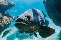
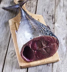
Species known to be a source of intoxication include barracudas, snappers, moray eels, parrotfish, groupers, triggerfish and amberjacks, but other species have been known to cause occasional outbreaks. Ciguatera toxins rarely contaminate pelagic fish such as tuna, marlins, dolphinfish or other ray-finned fish. Ciguatoxin can be found around the world in the tropical reef belt between 35 degrees north latitude and 35 degrees south latitude.
Epidemiology
Ciguatera is probably the most common type of marine food poisoning. It is endemic in Australia, the Caribbean and the South Pacific islands. Ciguatera cases should be naturally limited to these areas, but due to commercial imports, cases of ciguatera have been reported in areas like St. Louis, Missouri and New York City.
Approximately 50,000 reported cases of ciguatera poisoning occur annually worldwide. Epidemiological data regarding ciguatera poisoning is challenging to collect; because of the wide array of symptoms, ciguatera is often misdiagnosed or undiagnosed. People in endemic areas often disregard medical evaluation, while imported cases probably go undiagnosed or unreported, because physicians outside of endemic regions may be unfamiliar with symptoms of a tropical toxin. Recent studies have suggested that the incidence of this illness is continuing to increase, though this might be due to increased reporting rather than an increased occurrence of the disease.
Signs and Symptoms
Toxicity depends on exposure and dose (how much is ingested). Symptom onset usually occurs two to six hours after ingestion. Symptoms can last for weeks to years, and in some cases may lead to long-term disability.
Signs and symptoms can be highly variable, but typically include neurological or gastrointestinal manifestations; about 80 percent of patients showing varying degrees of impairment in both systems. The most common manifestations include:
- Gastrointestinal symptoms such as abdominal pain and gastroenteritis, nausea, vomiting or diarrhea. These initial symptoms typically resolve without intervention within a few hours.
- Neurological symptoms including paresthesia (tingling and numbness), ataxia (uncoordinated muscle movements) and vertigo. Severe cases may include cold allodynia (temperature reversal), a burning sensation upon contact with cold objects. Neurological symptoms may persist and are occasionally misdiagnosed as multiple sclerosis. In patients with a recent history of diving, muscular weakness and pain, these neurological symptoms can also be confounder for decompression illness.
- Skin itching that can persist for weeks and worsen as a result of activities that increase skin temperature like exercise and alcohol consumption.
Prevention
- Avoid consuming fish species commonly associated with ciguatera include barracuda, grouper, snapper, parrotfish, moray eels, triggerfish and amberjacks.
- Ciguatoxin is odorless, tasteless and heat-resistant—it will not taste different, and cooking will not prevent intoxication.
- While the whole fish will contain toxins, the highest concentrations are typically found in the liver, intestines and gonads.
Treatment
There is no definitive treatment for ciguatera poisoning. Both first aid and hospital care is aimed at symptom control. If vomiting is profuse, it is important to correct possible dehydration. If you suspect ciguatera, you should seek a medical evaluation. There are many folk remedies, but the efficacy of these has not been studied. The best course of action is prevention through education and avoidance of seafood in endemic or suspected areas.
The term ciguatera is actually inaccurate. “Ciguatera” was coined by Don Antonio Parra in Cuba in 1787 to describe an indigestion following ingestion of a type of marine snail called “cigua” (Turbo pica). The term “cigua” was somehow transferred to an intoxication caused by the ingestion of coral reef fish.
Scombroid Fish Poisoning
Scombroid fish poisoning is a foodborne illness that results from eating spoiled fish containing high amounts of histamine.
Source of Intoxication
There are many different species of fish that can be involved in scombroid poisoning, including mackerel, tuna, bonito, albacore, sardines, anchovies, mahi-mahi, amberjacks, marlin and herrings.
If scombroids are poorly refrigerated after being caught, the fish will begin to decompose, and bacteria from the fish’s gastrointestinal tract will invade its flesh. Many fish contain a significant amount of an amino acid called histidine in their flesh. When decomposition begins, the bacteria from the gastrointestinal tract breaks histidine down into histamine (a small nitrogen compound involved in regulation of immune reactions and inflammatory responses). While ingestion of histidine is harmless, ingestion of large quantities of histamine can mimic an allergic reaction.
Epidemiology
In the United States and Europe, scombroid fish poisoning accounts for up to 40 percent of seafood-borne illness outbreaks. Between 1998 and 2002, there were 167 reported outbreaks in the United States affecting 703 persons with no fatalities. Scombroid fish poisoning can happen anywhere in the world where susceptible fish are harvested. This poisoning is more common when consuming fish caught recreationally or from small-scale operations; it rarely occurs in highly regulated fish harvests.
Signs and Symptoms
Ingestion of large quantities of histamine can mimic an allergic reaction. Symptom onset may range from minutes after consumption to up to two hours and typically resolves within 24 hours.
Symptoms may include:
- Skin flushing
- Oral burning
- Nausea
- Abdominal cramps
- Diarrhea
- Palpitations
- Sweating
Signs may consist of:
- Redness (diffuse erythema)
- Elevated heart rate at rest (tachycardia)
- Hypo- or hypertension
- Wheezing (likely in individuals with a history of asthma, chronic obstructive pulmonary disease or reactive airway disease)
Due to its resemblance to an allergic reaction combined with poor knowledge of intoxication, scombroid fish poisoning is commonly misdiagnosed as a seafood allergy. Anyone showing signs and symptoms compatible with allergic reactions should seek an immediate medical evaluation as allergic and allergic-like reactions can be life threatening.
Prevention
- Scombroid fish poisoning is entirely preventable by immediately storing fresh fish in coolers or ice containers away from direct sunlight. The Centers for Disease Control and Prevention (CDC) recommends temperatures below 40°F (4.4°C) at all points during the fish supply chain.
- Affected fish may have a peppery taste, but normal taste does not guarantee safety.
- Histamine is heat stable, so cooking does not prevent scombroid fish poisoning.
Treatment
As opposed to genuine allergic reactions, where the source of histamine is internal, treatment for scombroid fish poisoning does not require the use of corticosteroids or adrenaline (epinephrine). Instead, scombroid fish poisoning responds very well to oral antihistamines, typically showing positive results within 10 to 15 minutes.
Never assume oral antihistamines are enough to control a presumed scombroid fish poisoning on your own. Always seek for professional medical evaluation and let a medical doctor decide over treatment and best course of action.
Red Tide & Shellfish Poisonings
Red tide is a colloquial term for a specific phenomenon known as harmful algal bloom. Occasionally, large concentrations of aquatic microorganisms naturally bloom in coastal areas. The rapid accumulation of algal blossom can be significant enough to cause a green, red or brown discoloration of estuarine and freshwater environments.
Scientists discourage the term red tide, because these phenomena are unrelated to tidal water movements and may not necessarily be red in color or present any discoloration at all. Instead, when these algal blooms are associated with potentially harmful toxins, a more precise and favored terminology is harmful algal bloom (HAB).
Negative Impact on Ecosystems
Among the involved microorganisms certain species of phytoplankton may be present, which can produce harmful natural toxins that can become concentrated in tissues of filter feeders like shellfish and other mollusks and crustaceans. The whole food chain may be affected, and millions of fish may die as a result.
Danger to Humans
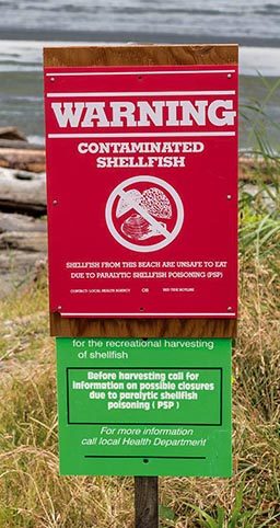
These toxins can affect commercial fisheries and represent a public health threat. People who consume contaminated shellfish may suffer a variety of shellfish poisonings, some of which are potentially lethal. Hazards related to HAB may not be limited to shellfish consumption, so avoid harvesting any type seafood on areas where HAB is known to have endemic outbreaks.
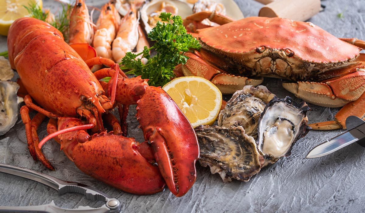
Shellfish Poisonings
Shellfish are bivalve (two-part shells) mollusks that capture nutrients by filtering water. During this process, these filter feeders can accumulate toxins and other contaminants. When humans consume these bivalves, they may be poisoned. These toxins are water-soluble and heat- and acid-stable—they are unaltered by ordinary cooking methods. Shellfish poisonings are a group of four different syndromes caused by eating bivalve mollusks contaminated with toxins produced by microscopic algae.
SYNDROMES
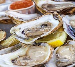
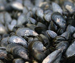
There are four different types of shellfish poisonings that are primarily associated with mollusks such as mussels, clams, oysters and scallops.
PARALYTIC SHELLFISH POISONING (PSP)
These mollusks can accumulate a toxin called saxitoxin, which is produced by phytoplankton (dinoflagellates, diatoms and cyanobacteria). Some shellfish remain toxic for several weeks, while others can store the toxin for up to two years.
PSP blooms are associated with harmful algal blooms, which can occur in almost all oceans. PSP can be fatal, particularly in children. Symptoms can appear a few minutes after ingestion and include nausea, vomiting, diarrhea, abdominal cramps, numbness or burning around the mouth, gums, tongue and progressing to the neck, arms, legs and toes. Other symptoms may include dry mouth, shortness of breath, slurred speech and loss of consciousness. Signs of toxicity and mortality are also seen in wild animals.
AMNESIC SHELLFISH POISONING (ASP)
This rare syndrome is caused by consuming shellfish contaminated with a toxin called domoic acid produced by certain marine diatoms.
Symptoms can appear 24 hours after ingestion of contaminated mollusks and may include nausea, vomiting, diarrhea, abdominal cramps and hemorrhagic gastritis. Neurological signs are severe and can take up to three days to develop. Neurological signs include dizziness, disorientation, visual disturbances, short-term memory loss, motor weakness, seizures, increased respiratory secretions and life-threatening dysrhythmias (irregular heartbeat). Death is rare. Resulting conditions due to permanent damage to the central nervous system may include short-term memory loss and peripheral neuropathy (weakness, numbness or pain as a result of nerve damage).
DIARRHEAL SHELLFISH POISONING (DSP)
Certain dinoflagellates produce a toxin known as okadaic acid that can cause a diarrheic syndrome. This toxin can damage the intestinal mucous membrane making it very permeable to water, which causes significant diarrhea as well as nausea, vomiting and abdominal cramps.
Symptoms can strike within a few minutes to an hour of ingesting contaminated shellfish and can last for about one day. No life-threatening symptoms have ever been recorded, but serious dehydration may occur.
NEUROTOXIC SHELLFISH POISONING (NSP)
NSP is caused by a toxin called brevetoxin, naturally produced by a dinoflagellate known as Karenia brevis. Brevetoxin can cause a variety of neurological symptoms very similar to ciguatera. NSP is generally not life threatening, but hospitalization is recommended until all other possible causes have been ruled out. In the United States and the Gulf of Mexico, a blossom of Karenia brevis usually causes the phenomena known as HAB.
Prevention
HABs occur throughout the world, killing millions of marine animals and affecting fisheries. Before harvesting your own seafood from coastal areas, research where HABs may occur and avoid consuming self-caught shellfish and fish from areas known to have HABs. Commercial fisheries tend to be safer than small scale artisanal harvesters.
The National Oceanic and Atmospheric Administration (NOAA) has a NOAA HAB (Red Tide) Watch page on Facebook. This system provides an operational forecast for harmful algal blooms. For those not on Facebook, NOAA’s Tides & Currents portal also provides an Operational Forecast System for HABs.
The Florida Fish and Wildlife Conservation Commission offers an online resource with a current map of Red Tide counts in the state of Florida.
Next: Chapter 4 – Appendix >